Mouse Glypican 6 Antibody
R&D Systems, part of Bio-Techne | Catalog # AF1053


Key Product Details
Species Reactivity
Validated:
Cited:
Applications
Validated:
Cited:
Label
Antibody Source
Product Specifications
Immunogen
Asp24-Val513
Accession # Q9R087
Specificity
Clonality
Host
Isotype
Scientific Data Images for Mouse Glypican 6 Antibody
Detection of Mouse Glypican 6 by Western Blot
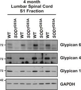
GDE2 substrates show impaired release in SOD1G93A mice. a Schematic of the sequential solubilization protocol used to separate hydrophilic non-membrane proteins (S1) from hydrophobic membrane proteins (S2). b Western blot demonstrating the successful enrichment of non-membrane protein (GAPDH) in the S1 fraction and membrane protein (Na/K ATPase) in the S2 fraction for WT and SOD1G93A transgenic mice. c Western blots visualizing the impaired production of non-membrane bound (S1) Glypican 6, Glypican 4, and Glypican 1 in the lumbar spinal cord of 4 month SOD1G93A mice. d Quantification of the reduced membrane release of Glypican 6, *p = 0.038; Glypican 4, *p = 0.016; and Glypican 1, *p = 0.030 relative to GAPDH. Graph represents mean ± SEM. Student’s t test, n = 4 WT, 4 SOD1G93A. Image collected and cropped by CiteAb from the following open publication (https://molecularneurodegeneration.biomedcentral.com/articles/10.1186/s13024-017-0148-1), licensed under a CC-BY license. Not internally tested by R&D Systems.Detection of Mouse Glypican 6 by Western Blot
GDE2 substrates show impaired release in SOD1G93A mice. a Schematic of the sequential solubilization protocol used to separate hydrophilic non-membrane proteins (S1) from hydrophobic membrane proteins (S2). b Western blot demonstrating the successful enrichment of non-membrane protein (GAPDH) in the S1 fraction and membrane protein (Na/K ATPase) in the S2 fraction for WT and SOD1G93A transgenic mice. c Western blots visualizing the impaired production of non-membrane bound (S1) Glypican 6, Glypican 4, and Glypican 1 in the lumbar spinal cord of 4 month SOD1G93A mice. d Quantification of the reduced membrane release of Glypican 6, *p = 0.038; Glypican 4, *p = 0.016; and Glypican 1, *p = 0.030 relative to GAPDH. Graph represents mean ± SEM. Student’s t test, n = 4 WT, 4 SOD1G93A Image collected and cropped by CiteAb from the following open publication (https://molecularneurodegeneration.biomedcentral.com/articles/10.1186/s13024-017-0148-1), licensed under a CC-BY license. Not internally tested by R&D Systems.Applications for Mouse Glypican 6 Antibody
Immunohistochemistry
Sample: Immersion fixed frozen sections of mouse embryo (E13)
Western Blot
Sample: Recombinant Mouse Glypican 6
Formulation, Preparation, and Storage
Purification
Reconstitution
Formulation
Shipping
Stability & Storage
- 12 months from date of receipt, -20 to -70 °C as supplied.
- 1 month, 2 to 8 °C under sterile conditions after reconstitution.
- 6 months, -20 to -70 °C under sterile conditions after reconstitution.
Background: Glypican 6
Glypican 6 (glypiated proteoglycan 6; also GPC6) is an 85-95 kDa heparan sulfate proteoglycan member of the GPI-linked glypican family of molecules. It is expressed by postnatal chrondrocytes and embryonic mesenchyme (connective tissue plus smooth muscle cells), and potentially modulates the availability, or signaling, of heparin-binding growth factors. Mouse Glypican 6 is synthesized as a 532 amino acid (aa) proprecursor that contains a 507 mature region (aa 4-530) and a 25 aa C-terminal prosegment. There is one potential splice variant that shows a 10 aa insertion after Leu384. Over aa 4-513, mouse Glypican 6 shares 99% aa identity with both human and rat Glypican 6.
Additional Glypican 6 Products
Product Documents for Mouse Glypican 6 Antibody
Product Specific Notices for Mouse Glypican 6 Antibody
For research use only
