Mouse IL-1 RI Antibody
R&D Systems, part of Bio-Techne | Catalog # AF771


Key Product Details
Species Reactivity
Validated:
Cited:
Applications
Validated:
Cited:
Label
Antibody Source
Product Specifications
Immunogen
Leu20-Lys338
Accession # P13504
Specificity
Clonality
Host
Isotype
Endotoxin Level
Scientific Data Images for Mouse IL-1 RI Antibody
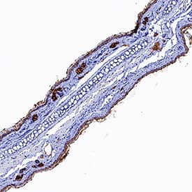
IL‑1 RI in Mouse Skin.
IL-1 RI was detected in perfusion fixed frozen sections of mouse skin (tail) using Goat Anti-Mouse IL-1 RI Antigen Affinity-purified Polyclonal Antibody (Catalog # AF771) at 1 µg/mL for 1 hour at room temperature followed by incubation with the Anti-Goat IgG VisUCyte™ HRP Polymer Antibody (Catalog # VC004). Tissue was stained using DAB (brown) and counterstained with hematoxylin (blue). Specific staining was localized to developing central nervous system. View our protocol for IHC Staining with VisUCyte HRP Polymer Detection Reagents.Cell Proliferation Induced by IL‑1 alpha/IL‑1F1 and Neutral-ization by Mouse IL‑1 RI Antibody.
Recombinant Mouse IL-1a/IL-1F1 (Catalog # 400-ML) stimulates proliferation in the the D10.G4.1 mouse helper T cell line in a dose-dependent manner (orange line). Proliferation elicited by Recombinant Mouse IL-1a/IL-1F1 (50 pg/mL) is neutralized (green line) by increasing concentrations of Goat Anti-Mouse IL-1 RI Antigen Affinity-purified Polyclonal Antibody (Catalog # AF771). The ND50 is typically 0.2-1.0 µg/mL.Applications for Mouse IL-1 RI Antibody
Immunohistochemistry
Sample: Perfusion fixed frozen sections of mouse lymph node and thymus, and mouse skin (tail)
Western Blot
Sample: Recombinant Mouse IL-1 RI Fc Chimera (Catalog # 771-MR)
Neutralization
Reviewed Applications
Read 5 reviews rated 2.6 using AF771 in the following applications:
Formulation, Preparation, and Storage
Purification
Reconstitution
Formulation
Shipping
Stability & Storage
- 12 months from date of receipt, -20 to -70 °C as supplied.
- 1 month, 2 to 8 °C under sterile conditions after reconstitution.
- 6 months, -20 to -70 °C under sterile conditions after reconstitution.
Background: IL-1 RI
Two distinct types of receptors that bind the pleiotropic cytokines IL-1 alpha and IL-1 beta have been described. The IL-1 receptor Type I is an 80 kDa transmembrane protein that is expressed predominantly by T cells, fibroblasts, and endothelial cells. IL-1 receptor Type II is a 68 kDa transmembrane protein found on B lymphocytes, neutrophils, monocytes, large granular leukocytes and endothelial cells. Both receptors are members of the immunoglobulin superfamily and show approximately 28% sequence identity in their extracellular domains. The two receptor types do not heterodimerize into a receptor complex. Mouse IL-1 RI shares 63% amino acid sequence homology with human IL-1 RI in their extracellular domains.
An IL-1 receptor accessory protein (1) that can heterodimerize with the Type I receptor in the presence of IL-1 alpha or IL-1 beta but not IL-1ra, was identified. This Type I receptor complex appears to mediate all the known IL-1 biological responses. The receptor Type II has a short cytoplasmic domain and does not transduce IL-1 signals. In addition to the membrane-bound form of IL-1 RII, a naturally-occurring soluble form of IL-1 RII has been described. It has been suggested that the Type II receptor, either as the membrane-bound or as the soluble form, serves as a decoy for IL-1 and inhibits IL-1 action by blocking the binding of IL-1 to the signaling Type I receptor complex. Recombinant IL-1 soluble receptor Type I is a potent antagonist of IL-1 action.
References
- Greenfeder, S. et al. (1995) J. Biol. Chem. 270:13757.
Long Name
Alternate Names
Gene Symbol
UniProt
Additional IL-1 RI Products
Product Documents for Mouse IL-1 RI Antibody
Product Specific Notices for Mouse IL-1 RI Antibody
For research use only
