Mouse IL-10 R alpha Antibody
R&D Systems, part of Bio-Techne | Catalog # AF-474-NA


Key Product Details
Validated by
Species Reactivity
Validated:
Cited:
Applications
Validated:
Cited:
Label
Antibody Source
Product Specifications
Immunogen
Thr24-Asn238 (Lys188Arg and Glu222Gly)
Accession # Q61727
Specificity
Clonality
Host
Isotype
Endotoxin Level
Scientific Data Images for Mouse IL-10 R alpha Antibody
Cell Proliferation Induced by IL-10 and Neutralization by Mouse IL-10 R alpha Antibody.
Recombinant Mouse IL-10 (Catalog # 417-ML) stimulates proliferation in the MC/9-2 mouse mast cell line in a dose-dependent manner (orange line). Proliferation elicited by Recombinant Mouse IL-10 (1 ng/mL) is neutralized (green line) by increasing concentrations of Goat Anti-Mouse IL-10 Ra Antigen Affinity-purified Polyclonal Antibody (Catalog # AF-474-NA). The ND50 is typically 1-3 µg/mL.Detection of Mouse IL-10R alpha by Immunocytochemistry/ Immunofluorescence
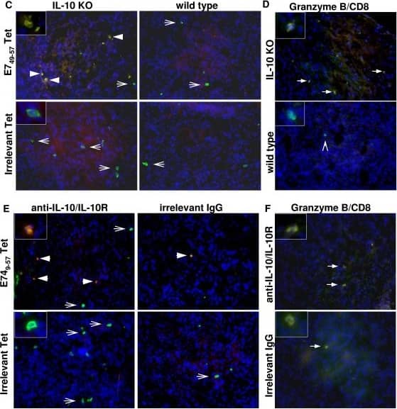
Lymphocyte infiltrate in tumors from IL-10 deficient mice (A, C and D) or from anti-IL10/IL-10R neutralizing antibody treated mice (B, E and F). A. Single cell suspensions were stained with the indicated antibodies plus anti-CD45 and anti-F4/80. Lymphocytes were analyzed in the CD45+F4/80- population. B. After 6 hours incubation in tissue culture flasks, non adherent cells from total tumor suspension were harvested and stained with the indicated antibodies, tetramer and anti-CD45. C and E. Immunofluorescence of cryo-sections from tumors harvested from IL-10 KO and wild type mice or neutralizing anti-IL10/IL-1R and irrelevant IgG antibodies, as indicated. Sections were stained with anti-CD8 (in FITC, green) and PE (red) conjugated HPV16 E749-57 tetramer - E749-57 Tet, and irrelevant tetramer - Irrelevant Tet. Cells were counterstained with DAPI (blue). Arrowheads indicate double positive cells in yellow, arrows indicate cells positive only for CD8. D and F. Granzyme B expression in tumor cryo-sections from IL-10 KO and wild type mice or mice treated with anti-IL10/IL-10R neutralizing antibodies and irrelevant IgG, as indicated. Sections were stained with anti-CD8 (green), followed by fixation, permeabilization and staining with anti-Granzyme B (red). Tissue was counterstained with DAPI. Solid arrows indicate double positive cells, arrowhead indicate CD8 only positive cell. Insets show details of cells positive only for CD8 (green) or double positive CD8/tetramer (C and E), CD8/Granzyme B (D and F) in yellow or orange due to the sobreposition of emission from FITC and PE (tetramer) or alexa 594 (Granzyme B). Image collected and cropped by CiteAb from the following open publication (https://bmcimmunol.biomedcentral.com/articles/10.1186/1471-2172-11-27), licensed under a CC-BY license. Not internally tested by R&D Systems.Detection of Porcine IL-10R alpha by Western Blot
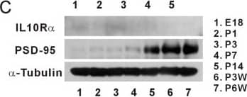
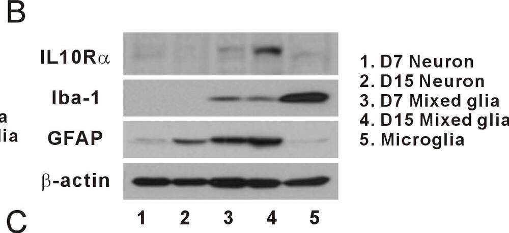
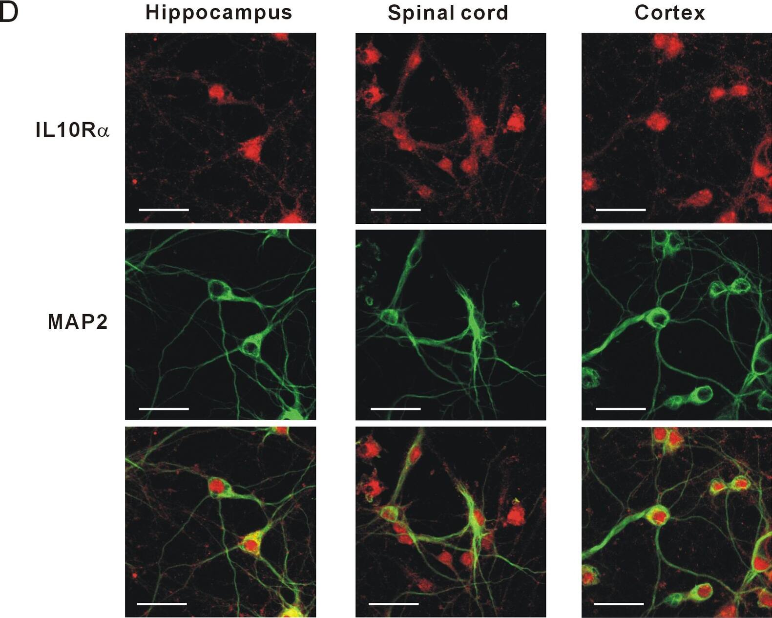
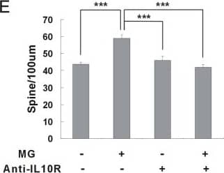
IL-10 receptors expressed on hippocampal neurons.(A) Expression of IL-10 receptor mRNAs in hippocampal neurons. The expression of the IL-10 receptor was identified using RT-PCR. The mRNAs of IL-10 receptor alpha and beta were expressed in the hippocampal neurons. IL-10 receptor alpha was expressed mainly in hippocampal neurons of DIV 7. (1, cultured hippocampal neurons at DIV 7; 2, cultured hippocampal neurons at DIV 15; 3, mixed glial culture at DIV 7; 4, mixed glial culture at DIV 15; 5, microglia) Quantification (DIV 15 neuron/ DIV 7 neuron): IL-10 receptor alpha, 0.61; IL-10 receptor beta, 1.06. (B) Expression of IL-10 receptor proteins in cultured hippocampal neurons. Similar to the expression of mRNA, the IL-10 receptor alpha protein was expressed mainly in neurons of DIV 7. Anti-IL-10 receptor alpha antibodies (0.5 μg/ml, Santa Cruz, sc-985) were used for western blotting [27]. Quantification (DIV 15 neuron/ DIV 7 neurons): IL-10 receptor alpha, 0.73.(C) Expression of IL-10 receptor proteins in the developing rat brains. The IL-10 receptor alpha proteins were expressed mainly in the developing brains of embryonic and early postnatal days (E18~P3). Quantification of IL-10 receptor alpha: E18, 0.30; P1, 0.27; P3, 1.0; P7, 0.22; P14, 0.20; P3W, 0.17; P6W, 0.15 (E, embryonic days; P, postnatal days).(D) Images of IL-10 receptor expressions in cultured hippocampal neurons. Hippocampal neurons of DIV 6 were stained with antibodies of IL-10 receptor alpha (5 μg/ml, Santa Cruz, sc-985) (red) and MAP2 (the neuronal marker, green) after treatment with 4% formaldehyde and then -20 °C methanol. Hippocampal neurons expressed IL-10 receptor proteins comparable to spinal neurons or cortical neurons.(E) The induction of synaptic formation by microglia was antagonized by the neutralizing antibody of IL-10 receptor alpha. When anti-mouse IL-10 receptor alpha antibody was applied to the co-culture of mouse microglia and mouse hippocampal neurons, the density of dendritic spines was significantly decreased compared with the control (without anti-IL-10 receptor antibody). Means±SEM. n=30 dendrites for no microglia without anti-IL-10 receptor antibody, 29 for the application of microglia only, 29 for no microglia with anti-IL-10 receptor antibody only, 29 for microglia with anti-IL-10 receptor antibody. ***p<0.001 by the Newman-Keuls multiple comparison test after application of one-way ANOVA, F=17.35, p<0.0001.(F) The induction of synaptic formation via microglia was not antagonized by the blocking antibody of TNF alpha. When anti-rat TNF alpha antibody was applied to the co-culture of rat microglia and rat hippocampal neurons, the density of dendritic spine was not decreased compared with control (without anti-TNF alpha antibody). Means±SEM. n=27 dendrites for no microglia without anti-TNF alpha antibody, 28 for the application of microglia only, 29 for no microglia with anti-TNF alpha receptor antibody only, 27 for microglia with anti-TNF alpha antibody. **p<0.01 and ***p<0.001 by the Newman-Keuls multiple comparison test after application of one-way ANOVA, F=9.104, p<0.0001. Image collected and cropped by CiteAb from the following open publication (https://pubmed.ncbi.nlm.nih.gov/24278397), licensed under a CC-BY license. Not internally tested by R&D Systems.Applications for Mouse IL-10 R alpha Antibody
Western Blot
Sample: Recombinant Mouse IL-10 R alpha (Catalog # 474-MR)
Neutralization
Formulation, Preparation, and Storage
Purification
Reconstitution
Formulation
Shipping
Stability & Storage
- 12 months from date of receipt, -20 to -70 °C as supplied.
- 1 month, 2 to 8 °C under sterile conditions after reconstitution.
- 6 months, -20 to -70 °C under sterile conditions after reconstitution.
Background: IL-10 R alpha
IL-10, initially designated cytokine synthesis inhibitory factor (CSIF), is a potent immunosuppressant of macrophage functions. IL-10 is also a pleiotropic cytokine with multiple immunostimulatory as well as immunosuppressive effects on a variety of other cell types. IL-10 binds specifically and with high affinity to cell-surface receptors. Mouse and human cDNA clones encoding the ligand-binding IL-10 receptor (IL-10 R) have been isolated. The IL-10 R mRNA has been detected in all cell types that are known to respond to IL-10.
Human and mouse IL-10 receptors are structurally related to the IFN-gamma receptor. These receptors are members of the class II subgroup of the cytokine receptor superfamily. The deduced amino acid sequence of human IL-10 R is approximately 60% identical to mouse IL-10 R. Although human IL-10 has cross-species activities and is active on mouse cells, mouse IL-10 is species-specific in its actions and does not bind to the human IL-10 receptor. The human IL-10 R gene has been mapped to chromosome 11q23.3. Recombinant IL-10 soluble receptor, consisting of the extracellular domain of IL-10 R, binds IL-10 with high affinity in solution and is a potent IL-10 antagonist.
Long Name
Alternate Names
Gene Symbol
UniProt
Additional IL-10 R alpha Products
Product Documents for Mouse IL-10 R alpha Antibody
Product Specific Notices for Mouse IL-10 R alpha Antibody
For research use only
