Mouse IL-18 BPc Antibody
R&D Systems, part of Bio-Techne | Catalog # AF129


Key Product Details
Validated by
Species Reactivity
Validated:
Cited:
Applications
Validated:
Cited:
Label
Antibody Source
Product Specifications
Immunogen
Arg25-Ser192
Accession # AAD17193
Specificity
Clonality
Host
Isotype
Scientific Data Images for Mouse IL-18 BPc Antibody
Detection of Mouse IL-18 BPc by Western Blot
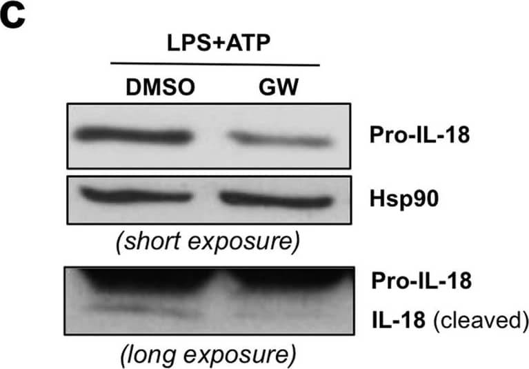
LPS-induced IL-18 levels are regulated by LXR activation.(a) BMDM were treated with vehicle (DMSO), GW3965 (1 μ mol/L) or LXR antagonist (LXR ant, 1 μ mol/L) for 24 hours with or without LPS (100 ng/ml) for the last 6 hours. IL-18 mRNA levels were analyzed by RT-qPCR. Values indicate expression normalized to cyclophilin and are presented relative to the expression in LPS-treated cells, set as 1. Values represent the mean ± SEM (3–4 different BMDM preparations). t-test: ***p ≤ 0.001, ns p > 0.05. (b) IL-18 mRNA expression in BMDM from WT and LXR alpha beta −/− mice were analyzed by RT-qPCR. Shown is a representative experiment performed in triplicate (mean ± SD) t-test: *p ≤ 0.05, ns p > 0.05. (c) BMDM were treated as in A and then activated with ATP for the last 2 hours. Pro-IL-18 expression was analyzed by immunoblotting and Hsp90 levels were assayed as loading control. Shown is a representative experiment of three. (d) BMDM were treated as in A. Intracellular expression of IL-18 is shown as analyzed by immunoblotting. Samples shown were analysed on non consecutive lanes of the same blot. (e) BMDM were treated as indicated. Secreted IL-18 levels in culture supernatants were quantified by ELISA. Values represent mean ± SEM (n = 3). t-test: *p ≤ 0.05. Image collected and cropped by CiteAb from the following open publication (https://www.nature.com/articles/srep25481), licensed under a CC-BY license. Not internally tested by R&D Systems.Detection of Mouse IL-18 BPc by Western Blot
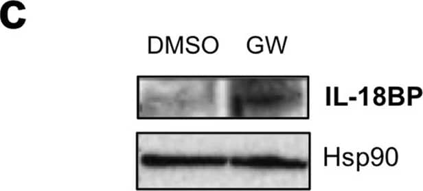
Identification of IL-18BP as a novel LXR target gene.(a) BMDM were treated with vehicle (DMSO) or GW3965 (GW) for 24 hours and IL-18BP mRNA content was analyzed by RT-qPCR. Values indicate expression normalized to cyclophilin and are presented relative to the expression in vehicle-treated cells. Values represent the mean of 4 independent experiments ± SEM. t-test: ***p ≤ 0.001. (b) BMDM from WT or LXR alpha beta deficient mice (LXR alpha beta −/−) were treated as in A with or without the RXR activator LG268 (LG). IL-18BP mRNA content was analyzed as in A. Values represent the mean of 3 independent experiments ± SEM. *p ≤ 0.05, **p ≤ 0.01. (c) BMDM were treated with vehicle (DMSO) or GW3965 (GW) for 24 hours supplemented with Brefeldin A for the last 6 hours. Protein content was analyzed by immunoblotting and Hsp90 levels were assayed as loading control. (d) RAW264.7 cells were transfected with hLXR alpha or pcDNA3.1 (− ) along with the indicated IL-18BP luciferase reporters or pGL3 empty vector. Cells were treated as described in the Methods section. For each reporter, luciferase and beta -galactosidase activities were measured and the ratio was compared to the vehicle-treated condition in the absence of LXR alpha (− ), which was set as 1. Data are mean value ± SD (n = 3) of one representative experiment. t-test: **p ≤ 0.01, ***p ≤ 0.001. (e) Upper panel, location of a putative LXRE in the IL-18BP locus. Arrows indicate the position of primers used for ChIP assays. Lower panel, BMDM cells were incubated with or without GW3965 at 1 μ mol/L for 2 hours. LXR occupancy was assessed by ChIP assays. Primers shown in upper panel were used to amplify an unrelated site at − 4.7 kb that served as a negative control, the − 1.1 kb putative LXRE, − 0.5 kb and TSS as GW3965 responsive sites. SREBP1c primers were used as a positive control for LXR binding. Shown is a representative experiment of three. Image collected and cropped by CiteAb from the following open publication (https://www.nature.com/articles/srep25481), licensed under a CC-BY license. Not internally tested by R&D Systems.Detection of Mouse IL-18 BPc by Western Blot
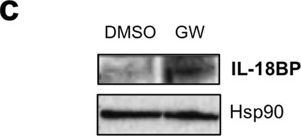
Identification of IL-18BP as a novel LXR target gene.(a) BMDM were treated with vehicle (DMSO) or GW3965 (GW) for 24 hours and IL-18BP mRNA content was analyzed by RT-qPCR. Values indicate expression normalized to cyclophilin and are presented relative to the expression in vehicle-treated cells. Values represent the mean of 4 independent experiments ± SEM. t-test: ***p ≤ 0.001. (b) BMDM from WT or LXR alpha beta deficient mice (LXR alpha beta −/−) were treated as in A with or without the RXR activator LG268 (LG). IL-18BP mRNA content was analyzed as in A. Values represent the mean of 3 independent experiments ± SEM. *p ≤ 0.05, **p ≤ 0.01. (c) BMDM were treated with vehicle (DMSO) or GW3965 (GW) for 24 hours supplemented with Brefeldin A for the last 6 hours. Protein content was analyzed by immunoblotting and Hsp90 levels were assayed as loading control. (d) RAW264.7 cells were transfected with hLXR alpha or pcDNA3.1 (− ) along with the indicated IL-18BP luciferase reporters or pGL3 empty vector. Cells were treated as described in the Methods section. For each reporter, luciferase and beta -galactosidase activities were measured and the ratio was compared to the vehicle-treated condition in the absence of LXR alpha (− ), which was set as 1. Data are mean value ± SD (n = 3) of one representative experiment. t-test: **p ≤ 0.01, ***p ≤ 0.001. (e) Upper panel, location of a putative LXRE in the IL-18BP locus. Arrows indicate the position of primers used for ChIP assays. Lower panel, BMDM cells were incubated with or without GW3965 at 1 μ mol/L for 2 hours. LXR occupancy was assessed by ChIP assays. Primers shown in upper panel were used to amplify an unrelated site at − 4.7 kb that served as a negative control, the − 1.1 kb putative LXRE, − 0.5 kb and TSS as GW3965 responsive sites. SREBP1c primers were used as a positive control for LXR binding. Shown is a representative experiment of three. Image collected and cropped by CiteAb from the following open publication (https://www.nature.com/articles/srep25481), licensed under a CC-BY license. Not internally tested by R&D Systems.Applications for Mouse IL-18 BPc Antibody
Western Blot
Sample: Recombinant Mouse IL-18 BPc
Formulation, Preparation, and Storage
Purification
Reconstitution
Formulation
Shipping
Stability & Storage
- 12 months from date of receipt, -20 to -70 °C as supplied.
- 1 month, 2 to 8 °C under sterile conditions after reconstitution.
- 6 months, -20 to -70 °C under sterile conditions after reconstitution.
Background: IL-18 BPc
Interleukin 18 binding protein (IL-18 BP) is a secreted glycoprotein, which functions as an IL-18 antagonist by binding to IL-18 and blocking its biological activity. IL‑18 BP bears no amino acid sequence homology to the membrane-associated IL-18 and IL-1 receptor proteins. The gene for human IL-18 BP has been localized to chromosome 11q13. It encodes for at least four isoforms by alternative splicing. The IL-18 BP isoforms a and c each contain one immunoglobulin (Ig)-like C2-type domain while isoforms b and d lack a complete Ig domain. The complete Ig domain has been shown to be essential to the binding and neutralizing properties of the binding proteins. Two isoforms of mouse IL18 BP (c and d) containing the complete Ig domain have also been isolated and shown to neutralize IL-18 bioactivity. Human and mouse IL-18 BPs share approximately 61% amino acid sequence identity. Several poxviruses also encode proteins with sequence similarity to the human and mouse IL-18 BP. Viral IL-18 BPs have been shown to bind and inhibit IL-18 responses and may be involved in modulating host immune responses. The expression of IL-18 BP is markedly up-regulated by IFN-gamma, suggesting that IL-18 activity is modulated by a negative feedback mechanism mediated by IL-18 BP.
Long Name
Alternate Names
Gene Symbol
UniProt
Additional IL-18 BPc Products
Product Documents for Mouse IL-18 BPc Antibody
Product Specific Notices for Mouse IL-18 BPc Antibody
For research use only

