Mouse IL-3 Antibody
R&D Systems, part of Bio-Techne | Catalog # MAB403

Key Product Details
Species Reactivity
Validated:
Cited:
Applications
Validated:
Cited:
Label
Antibody Source
Product Specifications
Immunogen
Accession # P01586
Specificity
Clonality
Host
Isotype
Endotoxin Level
Scientific Data Images for Mouse IL-3 Antibody
Cell Proliferation Induced by IL-3 and Neutralization by Mouse IL-3 Antibody.
Recombinant Mouse IL-3 (Catalog # 403-ML) stimulates proliferation in the NFS60 mouse myeloid cell line in a dose-dependent manner (orange line). Proliferation elicited by Recombinant Mouse IL-3 (0.5 ng/mL) is neutralized (green line) by increasing concentrations of Rat Anti-Mouse IL-3 Monoclonal Antibody (Catalog # MAB403). The ND50 is typically 0.05-0.15 µg/mL.Detection of Mouse IL-3 by Block/Neutralize
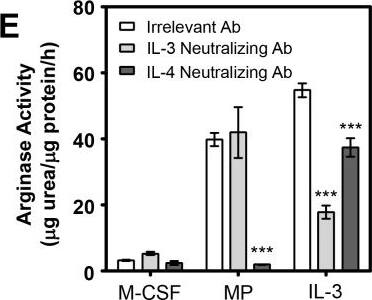
MP-induced M2 skewing of SHIP-/- BM progenitors requires IL-4 and basophils but not IL-3.A) Western blots of MФs derived from BM for 6 days with M-CSF ± IL-3 (10 ng/mL) or MP (5%). B) Arginase activity of MФs from SHIP-/- BM derived for 6 days with 10 ng/mL M-CSF ± IL-3 (10 ng/mL) or MP (5%) with 2.5 μg/mL of either an irrelevant or IL-4-neutralizing Ab. Data (mean ± SD) are representative of 3 independent experiments performed in triplicate. * p < 0.05 compared to all other conditions. C) Western blot of SHIP-/- MФs from (B) probed for Ym1, using SHC as a loading control. Dashed lines indicate where irrelevant lanes have been cropped out. All lanes are part of the same time-exposed film on the same gel. D) Western blot of SHIP-/- basophil-depleted (DX5-) or basophils (DX5+) + DX5- BM derived with M-CSF ± MP (5%). E) Arginase activity of SHIP-/- MФs derived from BM for 6 days with M-CSF ± MP (5%) or IL-3 (10 ng/mL) ± neutralizing Ab to IL-3 (2.5 μg/mL) or IL-4 (2.5 μg/mL). F) Western blot of cells corresponding to panel (E). *** p < 0.001 compared to relevant control. Image collected and cropped by CiteAb from the following publication (https://pubmed.ncbi.nlm.nih.gov/27977740), licensed under a CC-BY license. Not internally tested by R&D Systems.Applications for Mouse IL-3 Antibody
Western Blot
Sample: Recombinant Mouse IL-3 (Catalog # 403-ML)
Neutralization
Mouse IL-3 Sandwich Immunoassay
Reviewed Applications
Read 2 reviews rated 4 using MAB403 in the following applications:
Formulation, Preparation, and Storage
Purification
Reconstitution
Formulation
Shipping
Stability & Storage
- 12 months from date of receipt, -20 to -70 °C as supplied.
- 1 month, 2 to 8 °C under sterile conditions after reconstitution.
- 6 months, -20 to -70 °C under sterile conditions after reconstitution.
Background: IL-3
Interleukin 3 is a pleiotropic factor produced primarily by activated T cells that can stimulate the proliferation and differentiation of pluripotent hematopoietic stem cells as well as various lineage committed progenitors. In addition, IL-3 also affects the functional activity of mature mast cells, basophils, eosinophils and macrophages. Because of its multiple functions and targets, it was originally studied under different names, including mast cell growth factor P-cell stimulating factor, burst promoting activity, multi-colony stimulating factor, thy-1 inducing factor and WEHI-3 growth factor. In addition to activated T cells, other cell types such as human thymic epithelial cells, activated mouse mast cells, mouse keratinocytes and neurons/astrocytes can also produce IL-3. At the amino acid sequence level, mature human and mouse IL-3 share only 29% sequence identity. Consistent with this lack of homology, IL-3 activity is highly species-specific and human IL-3 does not show activity on mouse cells.
IL-3 exerts its biological activities through binding to specific cell surface receptors. The high affinity receptor responsible for IL-3 signaling is composed of alpha and beta subunits. The IL-3 R alpha is a member of the cytokine receptor super family and binds IL-3 with low affinity. Two distinct beta subunits, AIC2A ( betaIL-3) and AIC2B ( betac) are present in mouse cells. betaIL-3 also binds IL-3 with low affinity and forms a high affinity receptor with the alpha subunit. The betac subunits does not bind any cytokine but forms functional high affinity receptors with the alpha subunit of the IL-3, IL-5 and GM-CSF receptors. Receptors for IL-3 are present on bone marrow progenitors, macrophages, mast cells, eosinophils, megakaryocytes, basophils and various myeloid leukemic cells.
References
- Yokota, T. et al. (1984) Proc. Natl. Acad. Sci. USA 81:1070.
- Fung, M.C. et al. (1984) Nature 307:233.
- Miyatake, S. et al. (1985) Proc. Natl. Acad. Sci. USA 82:316.
Long Name
Alternate Names
Gene Symbol
UniProt
Additional IL-3 Products
Product Documents for Mouse IL-3 Antibody
Product Specific Notices for Mouse IL-3 Antibody
For research use only

