Mouse LIGHT/TNFSF14 Antibody
R&D Systems, part of Bio-Techne | Catalog # AF1794


Key Product Details
Species Reactivity
Validated:
Cited:
Applications
Validated:
Cited:
Label
Antibody Source
Product Specifications
Immunogen
Asp72-Val239
Accession # Q9QYH9
Specificity
Clonality
Host
Isotype
Scientific Data Images for Mouse LIGHT/TNFSF14 Antibody
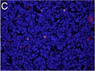
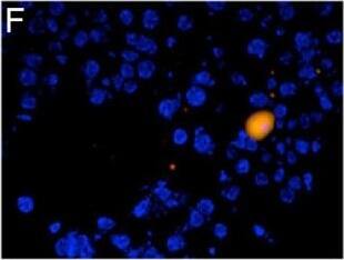
Detection of Mouse LIGHT/TNFSF14 by Immunocytochemistry/Immunofluorescence
Concurrent immunofluorescence staining of lymphocytes and LIGHT: Representative images of CD4+ staining (A,D red), LIGHT positive staining (B,E green) and co-expression (C,F yellow). DAPI was utilized as a nuclear counterstain (blue). Upper panels at low power identify an area of CD4+ TIL infiltration in a resected colorectal liver metastasis (200x). Lower panels demonstrate a single high power field in a peritumoral specimen (400x). Intratumoral CD3+ (14 ± 2.08 vs. 2.33 ± .67, p = .00006), CD4+ (8 ± 1.86 vs. 1.67 ± 0.33, p = .0009) and CD8+ (7 ± 2.33 vs. 2.0 ± 0.58, p = .029) lymphocytes were decreased in CRLM compared to lymphocytes from corresponding and equal areas of healthy control liver. Total CD3 + LIGHT + (0.33 ± 0.33 vs. 9 ± 2.65, p = .00006), CD4 + LIGHT + (0. 33 ± 0.33 vs. 5.33 ± .88, p = .0009) and CD8 + LIGHT + (1.33 ± .67 vs. 3.67 ± 1.33, p = .029) cells were decreased in tumor bearing liver compared to control. LIGHT-expressing CD3+ (6.33 ± 2.40 vs. 0.33 ± .33, p = .00006), CD4+ (5.33 ± 1.20 vs. 0.33 ± .33, p = .0009), and CD8+ (5.67 ± 2.40 vs. 1.67 ± 0.33, p = .029) lymphocytes were significantly higher in the peritumor region compared to the intratumor region (panels G-I)(n = 6). Image collected and cropped by CiteAb from the following publication (https://translational-medicine.biomedcentral.com/articles/10.1186/1479-5876-11-70), licensed under a CC-BY license. Not internally tested by R&D Systems.Detection of Mouse LIGHT/TNFSF14 by Immunocytochemistry/Immunofluorescence
Concurrent immunofluorescence staining of lymphocytes and LIGHT: Representative images of CD4+ staining (A,D red), LIGHT positive staining (B,E green) and co-expression (C,F yellow). DAPI was utilized as a nuclear counterstain (blue). Upper panels at low power identify an area of CD4+ TIL infiltration in a resected colorectal liver metastasis (200x). Lower panels demonstrate a single high power field in a peritumoral specimen (400x). Intratumoral CD3+ (14 ± 2.08 vs. 2.33 ± .67, p = .00006), CD4+ (8 ± 1.86 vs. 1.67 ± 0.33, p = .0009) and CD8+ (7 ± 2.33 vs. 2.0 ± 0.58, p = .029) lymphocytes were decreased in CRLM compared to lymphocytes from corresponding and equal areas of healthy control liver. Total CD3 + LIGHT + (0.33 ± 0.33 vs. 9 ± 2.65, p = .00006), CD4 + LIGHT + (0. 33 ± 0.33 vs. 5.33 ± .88, p = .0009) and CD8 + LIGHT + (1.33 ± .67 vs. 3.67 ± 1.33, p = .029) cells were decreased in tumor bearing liver compared to control. LIGHT-expressing CD3+ (6.33 ± 2.40 vs. 0.33 ± .33, p = .00006), CD4+ (5.33 ± 1.20 vs. 0.33 ± .33, p = .0009), and CD8+ (5.67 ± 2.40 vs. 1.67 ± 0.33, p = .029) lymphocytes were significantly higher in the peritumor region compared to the intratumor region (panels G-I)(n = 6). Image collected and cropped by CiteAb from the following publication (https://translational-medicine.biomedcentral.com/articles/10.1186/1479-5876-11-70), licensed under a CC-BY license. Not internally tested by R&D Systems.Applications for Mouse LIGHT/TNFSF14 Antibody
Western Blot
Sample: Recombinant Mouse LIGHT/TNFSF14 (Catalog # 1794-LT)
Reviewed Applications
Read 1 review rated 4 using AF1794 in the following applications:
Formulation, Preparation, and Storage
Purification
Reconstitution
Formulation
Shipping
Stability & Storage
- 12 months from date of receipt, -20 to -70 °C as supplied.
- 1 month, 2 to 8 °C under sterile conditions after reconstitution.
- 6 months, -20 to -70 °C under sterile conditions after reconstitution.
Background: LIGHT/TNFSF14
LIGHT (lymphotoxin-like, exhibits inducible expression, and competes with HSV glycoprotein D for HVEM, a receptor expressed by T lymphocytes) is a member of the TNF superfamily and is designated TNFSF14. The gene for mouse LIGHT encodes a 239 amino acid residue (aa) type II transmembrane glycoprotein that contains a 37 aa N-terminal cytoplasmic domain, a 21 aa transmembrane region, and a 181 aa extracellular domain. A soluble form of mouse LIGHT is generated from the membrane form by proteolytic processing. Similar to other TNF ligand family members, LIGHT is assembled as a homotrimer. Mouse and human LIGHT share 71% aa sequence identity.
LIGHT is expressed by activated lymphocytes, natural killer cells, immature dendritic cells, monocytes and granulocytes. Mouse LIGHT binds and signals via two distinct TNF receptor superfamily members, including the herpes virus entry mediator (HVEM/TNFRSF14) and the lymphotoxin beta receptor (LT beta R/TNFRSF3). In humans, LIGHT also binds the soluble human decoy receptor 3 (DcR3/TNFRSF6B). Signaling from LT beta R, which also binds LT alpha beta, induces apoptosis and the production of various cytokines. LIGHT-LT beta R signaling also plays a role in mesenteric lymph node organogenesis, and restoration of secondary lymphoid structure and function. Signaling from HVEM, which also binds LT alpha, co-stimulates T-helper cell type 1 (TH1) immune responses, enhances Cytotoxic T Lymphocytes (CTL)-mediated tumor immunity, and regulates allogeneic T cell activation and allograft rejection. Blockade of LIGHT-HVEM signaling has been shown to prevent graft versus host disease.
References
- Granger, S.W. and S. Rickert (2003) Cytokine Growth Factor Rev. 14:289.
Long Name
Alternate Names
Gene Symbol
UniProt
Additional LIGHT/TNFSF14 Products
Product Documents for Mouse LIGHT/TNFSF14 Antibody
Product Specific Notices for Mouse LIGHT/TNFSF14 Antibody
For research use only
