Mouse Lumican Antibody
R&D Systems, part of Bio-Techne | Catalog # AF2745

Key Product Details
Species Reactivity
Validated:
Mouse
Cited:
Human, Mouse
Applications
Validated:
Immunohistochemistry, Western Blot
Cited:
Immunocytochemistry, Immunohistochemistry, Immunohistochemistry-Paraffin, Immunoprecipitation, Western Blot
Label
Unconjugated
Antibody Source
Polyclonal Goat IgG
Product Specifications
Immunogen
Mouse myeloma cell line NS0-derived recombinant mouse Lumican
Gln19-Asn338, predicted
Accession # P51885
Gln19-Asn338, predicted
Accession # P51885
Specificity
Detects mouse Lumican in direct ELISAs and Western blots.
Clonality
Polyclonal
Host
Goat
Isotype
IgG
Scientific Data Images for Mouse Lumican Antibody
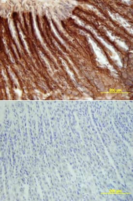
Lumican in Mouse Intestine.
Lumican was detected in perfusion fixed frozen sections of mouse intestine using Goat Anti-Mouse Lumican Antigen Affinity-purified Polyclonal Antibody (Catalog # AF2745) at 15 µg/mL overnight at 4 °C. Tissue was stained using the Anti-Goat HRP-DAB Cell & Tissue Staining Kit (brown; CTS008) and counterstained with hematoxylin (blue). Lower panel shows a lack of labeling if primary antibodies are omitted and tissue is stained only with secondary antibody followed by incubation with detection reagents. View our protocol for Chromogenic IHC Staining of Frozen Tissue Sections.Detection of Mouse Lumican by Immunohistochemistry
Immunoperoxidase for lumican. (A) Ovx group: A weak immunoreaction is observed in the endometrial stroma, especially under the luminal epithelium (arrow) and in the deep stroma, as well as in the EML and connective tissue between muscle layers of the myometrium; (B) E2-group: the reaction is present in the whole endometrial stroma and in both layers of the myometrium; (C) MPA-group: the reaction is present in the whole stroma, however it is weak in the subepithelial stroma (asterisk). In the myometrium, deposition of lumican is observed in the EML and connective tissue between layers; (D) E2+MPA-group: the immunoreaction is strong in the whole endometrial stroma. In the myometrium, a weak staining is observed in both IML and EML, being strong in the connective tissue between them. L = uterine lumen; G = endometrial gland; SS = superficial stroma; DS = deep stroma; V = blood vessel; IML = internal muscle layer; EML = external muscle layer. Scale bar: 50 μm. Image collected and cropped by CiteAb from the following publication (https://rbej.biomedcentral.com/articles/10.1186/1477-7827-9-22), licensed under a CC-BY license. Not internally tested by R&D Systems.Applications for Mouse Lumican Antibody
Application
Recommended Usage
Immunohistochemistry
5-15 µg/mL
Sample: Perfusion fixed frozen sections of mouse intestine, liver, lung, thymus, and spleen
Sample: Perfusion fixed frozen sections of mouse intestine, liver, lung, thymus, and spleen
Western Blot
0.1 µg/mL
Sample: Recombinant Mouse Lumican (Catalog # 2745-LU)
Sample: Recombinant Mouse Lumican (Catalog # 2745-LU)
Formulation, Preparation, and Storage
Purification
Antigen Affinity-purified
Reconstitution
Reconstitute at 0.2 mg/mL in sterile PBS. For liquid material, refer to CoA for concentration.
Formulation
Lyophilized from a 0.2 μm filtered solution in PBS with Trehalose. *Small pack size (SP) is supplied either lyophilized or as a 0.2 µm filtered solution in PBS.
Shipping
Lyophilized product is shipped at ambient temperature. Liquid small pack size (-SP) is shipped with polar packs. Upon receipt, store immediately at the temperature recommended below.
Stability & Storage
Use a manual defrost freezer and avoid repeated freeze-thaw cycles.
- 12 months from date of receipt, -20 to -70 °C as supplied.
- 1 month, 2 to 8 °C under sterile conditions after reconstitution.
- 6 months, -20 to -70 °C under sterile conditions after reconstitution.
Background: Lumican
References
- Nikitovic, D. et al. (2008) IUBMB Life 60:818.
- Blochberger, T.C. et al. (1992) J. Biol. Chem. 267:347.
- Chakravarti, S. et al. (1998) J. Cell Biol. 141:1277.
- Chakravarti, S. et al. (2000) Invest. Ophthalmol. Vis. Sci. 41:3365.
- Jepsen, K.J. et al. (2002) J. Biol. Chem. 277:35532.
- Chakravarti, S. et al. (2003) Invest. Ophthalmol. Vis. Sci. 44:2422.
- Vuillermoz, B. et al. (2004) Exp. Cell Res. 296:294
- Nikitovic, D. et al. (2008) FEBS J. 275:350.
- D’Onofrio, M.F. et al. (2008) Biochem. Biophys. Res. Commun. 365:266.
- Ishiwata, T. et al. (2007) Oncol. Rep. 18:537.
Alternate Names
LDC, LUM, SLRR2D
Gene Symbol
LUM
UniProt
Additional Lumican Products
Product Documents for Mouse Lumican Antibody
Product Specific Notices for Mouse Lumican Antibody
For research use only
Loading...
Loading...
Loading...
Loading...

