Mouse MAdCAM-1 Antibody
R&D Systems, part of Bio-Techne | Catalog # AF993


Key Product Details
Species Reactivity
Validated:
Cited:
Applications
Validated:
Cited:
Label
Antibody Source
Product Specifications
Immunogen
Gln22-Thr365, Predicted
Accession # NP_038619
Specificity
Clonality
Host
Isotype
Scientific Data Images for Mouse MAdCAM-1 Antibody
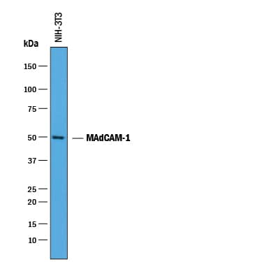
Detection of Mouse MAdCAM‑1 by Western Blot.
Western blot shows lysates of NIH-3T3 mouse embryonic fibroblast cell line. PVDF membrane was probed with 0.25 µg/mL of Goat Anti-Mouse MAdCAM-1 Antigen Affinity-purified Polyclonal Antibody (Catalog # AF993) followed by HRP-conjugated Anti-Goat IgG Secondary Antibody (Catalog # HAF019). A specific band was detected for MAdCAM-1 at approximately 50 kDa (as indicated). This experiment was conducted under reducing conditions and using Immunoblot Buffer Group 1.MAdCAM‑1 in Mouse Intestine.
MAdCAM-1 was detected in perfusion fixed frozen sections of mouse intestine using Goat Anti-Mouse MAdCAM-1 Antigen Affinity-purified Polyclonal Antibody (Catalog # AF993) at 1.7 µg/mL overnight at 4 °C. Tissue was stained using the Anti-Goat HRP-DAB Cell & Tissue Staining Kit (brown; Catalog # CTS008) and counterstained with hematoxylin (blue). Specific labeling was localized to the endothelial cells of blood capillaries in intestinal villi. View our protocol for Chromogenic IHC Staining of Frozen Tissue Sections.Applications for Mouse MAdCAM-1 Antibody
Immunohistochemistry
Sample: Perfusion fixed frozen sections of mouse intestine
Western Blot
Sample: NIH‑3T3 mouse embryonic fibroblast cell line
Reviewed Applications
Read 2 reviews rated 4 using AF993 in the following applications:
Formulation, Preparation, and Storage
Purification
Reconstitution
Formulation
Shipping
Stability & Storage
- 12 months from date of receipt, -20 to -70 °C as supplied.
- 1 month, 2 to 8 °C under sterile conditions after reconstitution.
- 6 months, -20 to -70 °C under sterile conditions after reconstitution.
Background: MAdCAM-1
Mucosal addressin cell adhesion molecule-1 (MAdCAM-1) is an immunoglobulin (Ig) cell adhesion molecule family member. In addition to Ig domains, it contains a mucin-like domain and a membrane proximal domain with similarity to IgA. MAdCAM-1 is involved in lymphocyte homing to mucosal sites and is expressed on high endothelial venules (HEV) of both mesenteric lymph nodes and Peyer’s patches. It has also been found to be expressed on sinus-lining cells of the spleen. The integrin, alpha4 beta7, has been shown to function as the MAdCAM-1 receptor. The Ig domains of MAdCAM-1 have been found to be critical to alpha4 beta7 binding. The mucin domain has been shown to have activity in L-Selectin binding. MAdCAM-1 expression has been demonstrated to be up-regulated by TNF-alpha and IL-1 beta. MAdCAM-1 appears to play a role in inflammatory bowel disease (IBD) as its expression is highly up-regulated in IBD and most likely serves to recruit alpha4 beta7-expressing lymphocytes to the region. In vivo studies involving nonobese diabetic (NOD) mice have also suggested that MAdCAM-1/ alpha4 beta7 interaction plays a role in diabetes development in this model. Mouse MAdCAM-1 is a 405 amino acid (aa) residue protein with a 21 aa signal sequence, a 344 aa extracellular domain, a 20 aa transmembrane domain and a 20 aa cytoplasmic domain.
References
- Briskin, M.J. et al. (1993) Nature 363:461.
- Yang, X.D. et al. (1997) Diabetes 46:1542.
- Sampaio, S.O. et al. (1995) J. Immunol. 155:2477.
- Kraal, G. et al. (1995) Am. J. Pathol. 147:763.
- Berg, E.L. et al. (1993) Nature 366:695.
- Takeuchi, M. and V.R. Baichwal (1995) Proc. Natl. Acad. Sci. USA 92:3561.
Long Name
Alternate Names
Gene Symbol
UniProt
Additional MAdCAM-1 Products
Product Documents for Mouse MAdCAM-1 Antibody
Product Specific Notices for Mouse MAdCAM-1 Antibody
For research use only
