Mouse Mer Antibody
R&D Systems, part of Bio-Techne | Catalog # MAB591

Key Product Details
Species Reactivity
Validated:
Cited:
Applications
Validated:
Cited:
Label
Antibody Source
Product Specifications
Immunogen
Glu23-Phe498
Accession # Q60805
Specificity
Clonality
Host
Isotype
Scientific Data Images for Mouse Mer Antibody
Mer in J774A.1 Mouse Cell Line.
Mer was detected in immersion fixed J774A.1 mouse reticulum cell sarcoma macrophage cell line using Rat Anti-Mouse Mer Monoclonal Antibody (Catalog # MAB591) at 10 µg/mL for 3 hours at room temperature. Cells were stained using the NorthernLights™ 557-conjugated Anti-Rat IgG Secondary Antibody (red; Catalog # NL013) and counterstained with DAPI (blue). Specific staining was localized to cell surfaces. View our protocol for Fluorescent ICC Staining of Non-adherent Cells.Detection of Mouse Mer by Western Blot
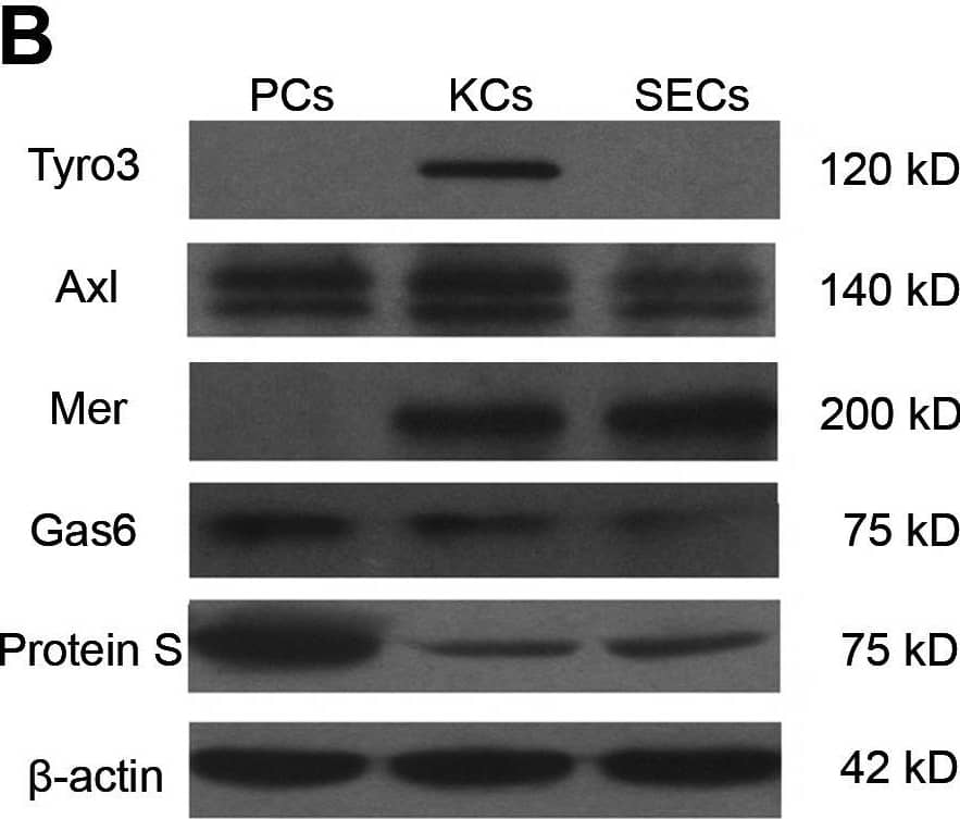
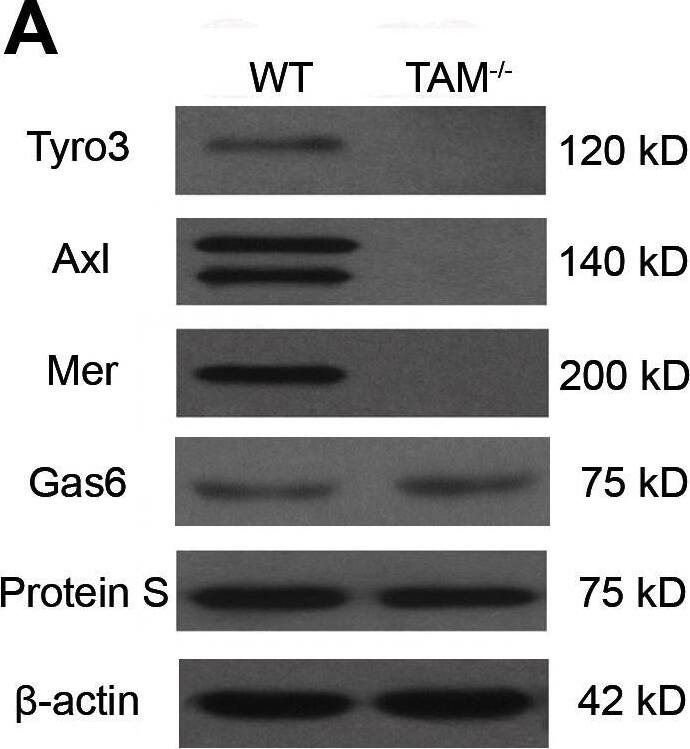
Expression of TAM RTKs, Gas6 and Protein S.(A) Western blot analyses of the liver lysates for the examination of TAM RTKs, Gas6 and Protein S. (B) Expression of TAM RTKs, Gas6 and Protein S in isolated liver cells: parenchymal cells (PCs), Kupffer cells(KCs) and sinusoidal endothelial cells (SECs). The primary cells were subjected to Western blotting. (C) Immunohistochemistry for the detection of TAM RTKs, Gas6 and Protein S. Arrowheads indicate PCs, and arrows indicate spindle-shaped sinusoidal cells corresponding to KCs and SECs. In negative controls (islets), sections were incubated with primary antibodies pre-incubated with an excess of blocking peptide. The livers of 15-week-old WT mice were used for the protein analyses. The images are representatives of at least three experiments. Scale bar = 20 µm. Image collected and cropped by CiteAb from the following open publication (https://pubmed.ncbi.nlm.nih.gov/23799121), licensed under a CC-BY license. Not internally tested by R&D Systems.Detection of Mouse Mer by Western Blot
Expression of TAM RTKs, Gas6 and Protein S.(A) Western blot analyses of the liver lysates for the examination of TAM RTKs, Gas6 and Protein S. (B) Expression of TAM RTKs, Gas6 and Protein S in isolated liver cells: parenchymal cells (PCs), Kupffer cells(KCs) and sinusoidal endothelial cells (SECs). The primary cells were subjected to Western blotting. (C) Immunohistochemistry for the detection of TAM RTKs, Gas6 and Protein S. Arrowheads indicate PCs, and arrows indicate spindle-shaped sinusoidal cells corresponding to KCs and SECs. In negative controls (islets), sections were incubated with primary antibodies pre-incubated with an excess of blocking peptide. The livers of 15-week-old WT mice were used for the protein analyses. The images are representatives of at least three experiments. Scale bar = 20 µm. Image collected and cropped by CiteAb from the following open publication (https://pubmed.ncbi.nlm.nih.gov/23799121), licensed under a CC-BY license. Not internally tested by R&D Systems.Applications for Mouse Mer Antibody
Immunocytochemistry
Sample: Immersion fixed J774A.1 mouse reticulum cell sarcoma macrophage cell line
Western Blot
Sample: Recombinant Mouse Mer Fc Chimera (Catalog # 591-MR)
Formulation, Preparation, and Storage
Purification
Reconstitution
Formulation
Shipping
Stability & Storage
- 12 months from date of receipt, -20 to -70 °C as supplied.
- 1 month, 2 to 8 °C under sterile conditions after reconstitution.
- 6 months, -20 to -70 °C under sterile conditions after reconstitution.
Background: Mer
Axl (Ufo, Ark), Dtk (Sky, Tyro3, Rse, Brt) and Mer (human and mouse homologues of chicken c-Eyk) constitute a receptor tyrosine kinase subfamily. The extracellular domains of these proteins contain two Ig-like motifs and two fibronectin type III motifs. This characteristic topology is also found in neural cell adhesion molecules and in receptor tyrosine phosphatases. These receptors bind the vitamin K-dependent protein growth-arrest-specific gene 6 (Gas6) which is structurally related to the anticoagulation factor protein S. Binding of Gas6 induces receptor autophosphorylation and downstream signaling pathways that can lead to cell proliferation, migration or the prevention of apoptosis. Studies suggest that this family of tyrosine kinase receptors may be involved in hematopoiesis, embryonic development, tumorigenesis and regulation of testicular functions (1-2).
References
- Nagata, K. et al. (1996) J. Biol. Chem. 22:30022.
- Crosier, K.E. and P.S Crosier (1997) Pathology 29:131.
Long Name
Alternate Names
Gene Symbol
UniProt
Additional Mer Products
Product Documents for Mouse Mer Antibody
Product Specific Notices for Mouse Mer Antibody
For research use only


