Mouse MFG-E8 Antibody
R&D Systems, part of Bio-Techne | Catalog # AF2805

Key Product Details
Validated by
Biological Validation
Species Reactivity
Validated:
Mouse
Cited:
Mouse, Bovine, Transgenic Mouse
Applications
Validated:
Simple Western, Western Blot
Cited:
Immunohistochemistry, Immunoprecipitation, In vivo assay, Western Blot
Label
Unconjugated
Antibody Source
Polyclonal Goat IgG
Product Specifications
Immunogen
Mouse myeloma cell line NS0-derived recombinant mouse MFG-E8 (R&D Systems, Catalog # 2805-MF)
Ala23-Cys463
Accession # P21956
Ala23-Cys463
Accession # P21956
Specificity
Detects mouse MFG-E8 in direct ELISAs and Western blots. In these formats, less than 2% cross‑reactivity with recombinant human MFG-E8 is observed.
Clonality
Polyclonal
Host
Goat
Isotype
IgG
Scientific Data Images for Mouse MFG-E8 Antibody
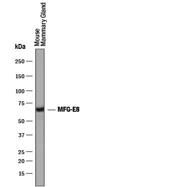
Detection of Mouse MFG-E8 by Western Blot.
Western blot shows lysates of mouse mammary gland tissue. PVDF membrane was probed with 0.25 µg/mL of Goat Anti-Mouse MFG-E8 Antigen Affinity-purified Polyclonal Antibody (Catalog # AF2805) followed by HRP-conjugated Anti-Goat IgG Secondary Antibody (Catalog # HAF019). A specific band was detected for MFG-E8 at approximately 60-70 kDa (as indicated). This experiment was conducted under reducing conditions and using Immunoblot Buffer Group 1.Detection of Mouse MFG‑E8 by Simple WesternTM.
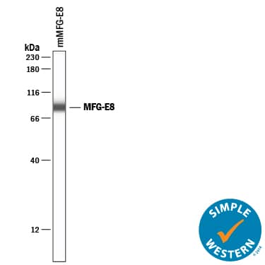
Simple Western lane view shows lysates of recombinant mouse (rm) MFG-E8 , loaded at 50 ng/mL. A specific band was detected for MFG-E8 at approximately 87 kDa (as indicated) using 50 µg/mL of Goat Anti-Mouse MFG-E8 Antigen Affinity-purified Polyclonal Antibody (Catalog # AF2805) followed by 1:50 dilution of HRP-conjugated Anti-Goat IgG Secondary Antibody (Catalog # HAF019). This experiment was conducted under reducing conditions and using the 12-230 kDa separation system.Detection of Human MFG-E8 by Western Blot
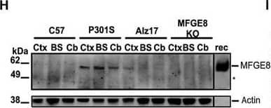
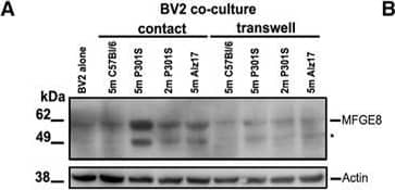
MFGE8 and NO Are Produced and Secreted by Co-cultured BV2 Microglia Only When Co-cultured in Contact with pFTAA+Neurons(A and B) Representative blot (A) ofMFGE8 in BV2 cells lysates cultured alone or co-cultured in direct contact, or via atranswell, with DRG neurons from 5-month-old P301S-tau, 5-month-old C57, 5-month-old Alz17 mice, or 2-month-old P301S mice.Asterisk: either another isoform ofMFGE8 or a breakdown product. (B) Densitometry of MFGE8 expression normalized to beta-actin; significantly more MFGE8 is produced only in contact co-cultures containing pFTAA+ neurons. Mean± SD, n = 3 independent experiments (*p < 0.05), twoway ANOVA, Dunnett’s correction.(C and D) Elevated MFGE8 (C) or NO (D) in medium conditioned by BV2 cells co-cultured in contact with DRG neurons from5-month-old P301S mice compared with 5-month-old Alz17, 5-month- old C57, or 2-month-old P301S mice (MFGE8: ****p < 0.0001versus all; NO: ****p < 0.0001 versus 5-month-old C57 or 2-month-old P301S mice; ***p < 0.001 versus 5-month-oldAlz17 mice). N.D., none detected. Mean ± SD, n = 3 independent experiments, two-way ANOVA, Bonferroni corrected.(E and F) Elevated MFGE8 (E) or NO (F) in medium conditioned by primary microglia from C57 mice co-cultured in contactwith DRG neurons from 5-month-old P301S mice; microglia from MFGE8 KO mice are negative controls. Mean ± SD, n = 3independent microglial preparations, one-way ANOVA, **p < 0.01 (MFGE8), *p < 0.05 (NO).(G) Increased MFGE8 immunostaining intensity in frontal motor cortex of 5-month-old P301S mice compared with 5-month-oldC57 mice; MFGE8 KO mouse is negative control; 25 μm section at inter- aural 5.12 mm, bregma 1.32 mm; brown, DAB; blue,cresyl violet. Scale bar, 130 mm. Inset, 65 μm.(H and I) Elevated MFGE8 (H) in 5-month-old P301S-tau brains. Lysatesfrom cortex(Ctx), brain stem (BS), and cerebellum(Cb) of 5-month-old C57, 5-month-old P301S-tau, and 5-month-old Alz17 mice probed with anti-MFGE8; MFGE8 KO brain lysate isnegative control. rec, recombinant mouse MFGE8. (I) Densitometry of MFGE8 expression normalized to beta-actin. Significantlyhigher MFGE8 expression in P301S versus C57BU6 Ctx (**p < 0.01), BS (****p < 0.0001), Cb (**p < 0.01), andP301SversusALz17BS (****p < 0.0001). Mean ± SD, n = 3 independent preparations, two-way ANOVA, Bonferronicorrected.(J) Elevated MFGE8 expression in brain extracts from cortex (Ctx) of FTDP-17T patients with two differentMAPT mutations (P301L, +3) or sporadic Pick’s disease but not in extracts from patients with C9orf72hexanucleotide expansions and TDP-43 aggregates. No expression is found in the cerebellum, where tau pathology is absent in allcases. Human milk MFGE8 is a positive control. Image collected and cropped by CiteAb from the following publication (https://pubmed.ncbi.nlm.nih.gov/30134156), licensed under a CC-BY license. Not internally tested by R&D Systems.Applications for Mouse MFG-E8 Antibody
Application
Recommended Usage
Simple Western
50 µg/mL
Sample: Recombinant Mouse (rm) MFG-E8
Sample: Recombinant Mouse (rm) MFG-E8
Western Blot
0.25 µg/mL
Sample: Mouse mammary gland tissue
Sample: Mouse mammary gland tissue
Reviewed Applications
Read 3 reviews rated 5 using AF2805 in the following applications:
Formulation, Preparation, and Storage
Purification
Antigen Affinity-purified
Reconstitution
Reconstitute at 0.2 mg/mL in sterile PBS. For liquid material, refer to CoA for concentration.
Formulation
Lyophilized from a 0.2 μm filtered solution in PBS with Trehalose. *Small pack size (SP) is supplied either lyophilized or as a 0.2 µm filtered solution in PBS.
Shipping
Lyophilized product is shipped at ambient temperature. Liquid small pack size (-SP) is shipped with polar packs. Upon receipt, store immediately at the temperature recommended below.
Stability & Storage
Use a manual defrost freezer and avoid repeated freeze-thaw cycles.
- 12 months from date of receipt, -20 to -70 °C as supplied.
- 1 month, 2 to 8 °C under sterile conditions after reconstitution.
- 6 months, -20 to -70 °C under sterile conditions after reconstitution.
Background: MFG-E8
References
- Raymond, A. et al. (2009) J. Cell. Biochem. 106:957.
- Stubbs, J.D. et al. (1990) Proc. Natl. Acad. Sci. USA 87:8417.
- Hoffhines, A.J. et al. (2008) J. Biol. Chem. 284:3096.
- Silvestre, J.-S. et al. (2005) Nat. Med. 11:499.
- Borges, E. et al. (2000) J. Biol. Chem. 275:39867.
- Hanayama, R. et al. (2002) Nature 417:182.
- Ait-Oufella, H. et al. (2007) Circulation 115:2168.
- Kranich, J. et al. (2010) J. Exp. Med. 207:2271.
- Hanayama, R. et al. (2004) Science 304:1147.
- Kranich, J. et al. (2010) J. Exp. Med. 205:1293.
- Atabai, K. et al. (2009) J. Clin. Invest. 119:3713.
- Bu, H.-F. et al. (2007) J. Clin. Invest. 117:3673.
- Jinushi, M. et al. (2009) J. Exp. Med. 206:1317.
- Kvistgaard, A.S. et al. (2004) J. Dairy Sci. 87:4088.
Long Name
Milk Fat Globule EGF Factor 8
Alternate Names
BA46, Breast epithelial antigen BA46, Lactahedrin, Medin, MFGE8, SED1
Gene Symbol
MFGE8
UniProt
Additional MFG-E8 Products
Product Documents for Mouse MFG-E8 Antibody
Product Specific Notices for Mouse MFG-E8 Antibody
For research use only
Loading...
Loading...
Loading...
Loading...
