Mouse PLA2G2A Antibody
R&D Systems, part of Bio-Techne | Catalog # AF4925

Key Product Details
Species Reactivity
Applications
Label
Antibody Source
Product Specifications
Immunogen
Asn22-Cys146
Accession # NP_001076000
Specificity
Clonality
Host
Isotype
Scientific Data Images for Mouse PLA2G2A Antibody
Detection of Mouse PLA2G2A by Western Blot.
Western blot shows lysates of mouse small intestine tissue. PVDF Membrane was probed with 0.5 µg/mL of Sheep Anti-Mouse PLA2G2A Antigen Affinity-purified Polyclonal Antibody (Catalog # AF4925) followed by HRP-conjugated Anti-Sheep IgG Secondary Antibody (Catalog # HAF016). A specific band was detected for PLA2G2A at approximately 17 kDa (as indicated). This experiment was conducted under reducing conditions and using Immunoblot Buffer Group 1.PLA2G2A in Mouse Intestine.
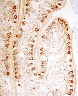
PLA2G2A was detected in perfusion fixed frozen sections of mouse intestine using Sheep Anti-Mouse PLA2G2A Antigen Affinity-purified Polyclonal Antibody (Catalog # AF4925) at 1.7 µg/mL overnight at 4 °C. Tissue was stained using the Anti-Sheep HRP-DAB Cell & Tissue Staining Kit (brown; Catalog # CTS019) and counterstained with hematoxylin (blue). Specific staining was localized to the cytoplasm of epithelial cells in intestinal glands. View our protocol for Chromogenic IHC Staining of Paraffin-embedded Tissue Sections.Detection of Mouse PLA2G2A by Simple WesternTM.
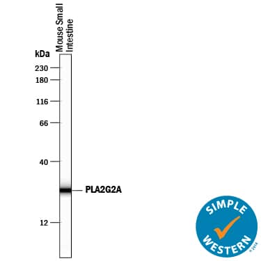
Simple Western lane view shows lysates of mouse small intestine tissue, loaded at 0.2 mg/mL. A specific band was detected for PLA2G2A at approximately 27 kDa (as indicated) using 5 µg/mL of Sheep Anti-Mouse PLA2G2A Antigen Affinity-purified Polyclonal Antibody (Catalog # AF4925) followed by 1:50 dilution of HRP-conjugated Anti-Sheep IgG Secondary Antibody (Catalog # HAF016). This experiment was conducted under reducing conditions and using the 12-230 kDa separation system.Applications for Mouse PLA2G2A Antibody
Immunohistochemistry
Sample: Perfusion fixed frozen sections of mouse intestine
Simple Western
Sample: Mouse small intestine tissue
Western Blot
Sample: Mouse small intestine tissue
Formulation, Preparation, and Storage
Purification
Reconstitution
Formulation
Shipping
Stability & Storage
- 12 months from date of receipt, -20 to -70 °C as supplied.
- 1 month, 2 to 8 °C under sterile conditions after reconstitution.
- 6 months, -20 to -70 °C under sterile conditions after reconstitution.
Background: PLA2G2A
Secretory Phospholipase A2 is an enzyme that hydrolyses the sn-2 ester bond of phospholipids, generating non-esterified free fatty acids and lysophospholipids (1‑3). PLA2G2A is a calcium-dependent phospholipase expressed in many cell types participating in inflammation-associated cellular responses, including platelets, neutrophils, and mast cells. It may function as an enzymatic component of the host defense mechanism. For example, human tears contain a high concentration of PLA2G2A, a principal bactericidal factor against Gram-positive bacteria in this fluid. It may play a role in cell proliferation through binding a receptor on the cell membrane. PLA2G2A has been shown to have pro-atherogenic properties both in the circulation and within the arterial wall (4). It is an acute phase protein expressed in response to a variety of pro-inflammatory cytokines. Circulating levels of sPLA2G2A are higher in coronary artery disease (CAD) patients and are associated with increased risk of future CAD (5).
References
- Webb, N. R. (2005) Cur. Opin. Lipid. 16:341.
- Triggiani, M. et al. (2005) J. Allergy Clin. Immunol. 116:1000.
- Murakami, M. and Kudo, I. (2004) Biol. Pharm. Bull. 27:1158.
- de Beer, F. C. and Webb, N. R. (2006) Arterioscler. Thromb. Vasc. Biol. 26:1421.
- Wootton, P. T. E. et al. (2006) Human Mol. Genet. 15:355.
Long Name
Alternate Names
Gene Symbol
UniProt
Additional PLA2G2A Products
Product Documents for Mouse PLA2G2A Antibody
Product Specific Notices for Mouse PLA2G2A Antibody
For research use only


