Mouse PRDM14 Antibody
R&D Systems, part of Bio-Techne | Catalog # MAB8097

Key Product Details
Species Reactivity
Validated:
Cited:
Applications
Validated:
Cited:
Label
Antibody Source
Product Specifications
Immunogen
Met1-Glu191
Accession # E9Q3T6
Specificity
Clonality
Host
Isotype
Scientific Data Images for Mouse PRDM14 Antibody
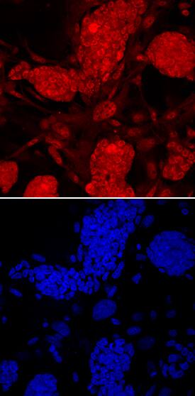
PRDM14 in D3 Mouse Embryonic Stem Cells.
PRDM14 was detected in immersion fixed D3 mouse embryonic stem cells using Rat Anti-Mouse PRDM14 Monoclonal Antibody (Catalog # MAB8097) at 10 µg/mL for 3 hours at room temperature. Cells were stained using the NorthernLights™ 557-conjugated Anti-Rat IgG Secondary Antibody (red; Catalog # NL013) and counterstained with DAPI (blue). Specific staining was localized to nuclei. View our protocol for Fluorescent ICC Staining of Stem Cells on Coverslips.Detection of Mouse PRDM14 by Western Blot
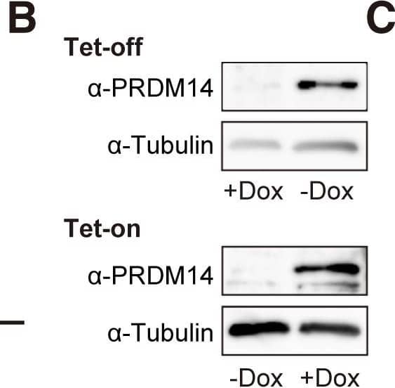
PRDM14 Induces the Transition of EpiLCs to ESCLCs(A) Constructs of the ROSA-Tet-off system and piggyBac Tet-on system.(B) Western blotting of PRDM14 and tubulin.(C) Scheme of the transition of ESCs to EpiLCs and of EpiLCs to ESCLCs.(D) qRT-PCR analysis of Prdm14 expression in ESCs, EpiLCs (d2), EpiLCs (d4), and EpiLCs + PRDM14. Error bars indicate ±SD of a biological triplicate.(E) AP staining of ESCs, EpiLCs (d2), EpiLCs (d4), and EpiLCs + PRDM14. Scale bar, 50 μm.(F) qRT-PCR analysis of pluripotency genes in ESCs, EpiLCs (d2), EpiLCs (d4), and EpiLCs + PRDM14. Error bars indicate ±SD of a biological triplicate.(G) Colony formation by ESCs, EpiLCs (d4), and EpiLCs + PRDM14 was assessed by AP staining over 3 days in the presence of serum + LIF. Error bars indicate ±SD of biological triplicates. p Values were calculated with the Student's t test. Scale bar, 200 μm.(H) (Left) Teratoma formation by EpiLCs expressing Prdm14 cultured in the absence of LIF. (Right) Immunofluorescent staining of alpha-fetoprotein (AFP), smooth muscle actin (SMA), and class III beta-tubulin (TUJ1) in teratoma sections. Scale bar, 200 μm.∗∗p < 0.01, ∗∗∗p < 0.001, Student's t test. Image collected and cropped by CiteAb from the following publication (https://pubmed.ncbi.nlm.nih.gov/27866876), licensed under a CC-BY license. Not internally tested by R&D Systems.Applications for Mouse PRDM14 Antibody
Immunocytochemistry
Sample: Immersion fixed D3 mouse embryonic stem cells
Formulation, Preparation, and Storage
Purification
Reconstitution
Formulation
Shipping
Stability & Storage
- 12 months from date of receipt, -20 to -70 °C as supplied.
- 1 month, 2 to 8 °C under sterile conditions after reconstitution.
- 6 months, -20 to -70 °C under sterile conditions after reconstitution.
Background: PRDM14
PRDM14 is an approximately 65 kDa transcriptional inhibitor that contains one histone methylating PR/SET domain (aa 243 – 360) and six zinc finger repeats (aa 390 - 558). PRDM14 is preferentially expressed in germ cells and undifferentiated embryonic stem cells in which it plays a central role in lineage specification, maintenance of pluripotency, and repression of somatic gene transcription. PRDM14 is upregulated in breast cancer and lymphoblastic leukemia. Within aa 1-191, mouse PRDM14 shares 46% aa sequence identity with human PRDM14.
Long Name
Alternate Names
Gene Symbol
UniProt
Additional PRDM14 Products
Product Documents for Mouse PRDM14 Antibody
Product Specific Notices for Mouse PRDM14 Antibody
For research use only

