Mouse R-Spondin 2 Antibody
R&D Systems, part of Bio-Techne | Catalog # MAB32661

Key Product Details
Species Reactivity
Validated:
Cited:
Applications
Validated:
Cited:
Label
Antibody Source
Product Specifications
Immunogen
Met1-Gly205
Accession # Q8BFU0
Specificity
Clonality
Host
Isotype
Endotoxin Level
Scientific Data Images for Mouse R-Spondin 2 Antibody
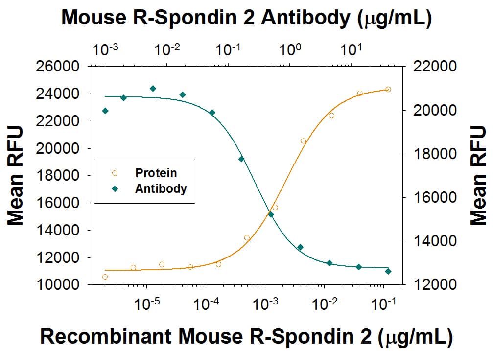
Β-Catenin Response Induced by R‑Spondin 2 and Neutralization by Mouse R‑Spondin 2 Antibody.
Recombinant Recombinant Mouse R-Spondin 2 (Catalog # 6946-RS) co-stimulates beta-catenin response in the HEK293T human embryonic kidney cell line in a dose-dependent manner (orange line). beta-Catenin response elicited by Mouse R-Spondin 2 (40 ng/mL) is neutralized (green line) by increasing concentrations of Rat Anti-Mouse R-Spondin 2 Monoclonal Antibody (Catalog # MAB32661) in the presence of 5 ng/mL of Recombinant Mouse Wnt-3a (Catalog # 1324-WN). The ND50 is typically 0.1-1 µg/ml.Applications for Mouse R-Spondin 2 Antibody
Neutralization
Formulation, Preparation, and Storage
Purification
Reconstitution
Formulation
Shipping
Stability & Storage
- 12 months from date of receipt, -20 to -70 °C as supplied.
- 1 month, 2 to 8 °C under sterile conditions after reconstitution.
- 6 months, -20 to -70 °C under sterile conditions after reconstitution.
Background: R-Spondin 2
Roof plate-specific Spondin 2 isoform 1 (R‑Spondin 2, RSPO2), also known as cysteine‑rich and single thrombospondin domain containing protein 2 (Cristin 2), is a 33 kDa secreted protein that belongs to the R‑Spondin family (1‑3). The four R‑Spondins regulate Wnt/ beta-catenin signaling and overlap in expression and function (1‑3). Like other R‑Spondins, RSPO2 contains two adjacent cysteine‑rich furin-like domains (aa 90‑134) followed by a thrombospondin (TSP-1) motif (aa 144‑204) and a C-terminal region rich in basic residues (aa 207‑243). The basic region binds heparin and mediates cell surface retention and extracellular matrix attachment while the furin-like domains are required for Wnt/ beta-catenin signaling (1, 3, 4). RSPO2 contains one potential N‑glycosylation site. Mature mouse RSPO2 shares 97‑98% aa identity with human, rat, equine, canine and bovine RSPO2 and ~40% aa identity with RSPO1, RSPO3 and RSPO4. One potential 237 aa mouse isoform diverges after aa 206 and lacks a basic region (5). Human RSPO2 is expressed in organs of endodermal origin in adults, including intestine and lung, and is down‑regulated in tumors of these tissues (1). In the embryonic mouse, RSPO2 expression is concentrated in the apical epidermal ridge, hippocampus, and developing muscle, teeth and bones (1, 6). Deletion of RSPO2 results in down‑regulation of Wnt activity in these areas, malformations of the facial skeleton and limbs, and respiratory failure at birth (7-9). RSPO2 is an extracellular potentiator of Wnt/ beta-catenin signaling (3, 4). It functions at least in part by binding LRP-6, stimulating its long-term phosphorylation and down‑regulating its internalization (3, 4). RSPO proteins, especially RSPO2 and RSPO3, also antagonize DKK1 activity by interfering with DKK1‑mediated LRP-6 and Kremen association (10).
References
- Kazanskaya, O. et al. (2004) Dev. Cell 7:525.
- Kim, K.-A. et al. (2006) Cell Cycle 5:23.
- Nam, J.-S. et al. (2006) J. Biol. Chem. 281:13247.
- Li, S-J. et al. (2009) Cell Signal. 21:916.
- Entrez accession # EDL08755.
- Nam, J.-S. et al. (2007) Gene Expr. Patterns 7:306.
- Yamada, W. et al. (2009) Biochem. Biophys. Res. Commun. 381:453.
- Jin, Y.-R. et al. (2011) Dev. Biol. 352:1.
- Nam, J.-S. et al. (2007) Dev. Biol. 311:124.
- Kim, K.-A. et al. (2007) Mol. Biol. Cell 19:2588.
Long Name
Alternate Names
Gene Symbol
UniProt
Additional R-Spondin 2 Products
Product Documents for Mouse R-Spondin 2 Antibody
Product Specific Notices for Mouse R-Spondin 2 Antibody
For research use only
