Mouse/Rat Cathepsin L Antibody
R&D Systems, part of Bio-Techne | Catalog # AF1515


Key Product Details
Validated by
Species Reactivity
Validated:
Cited:
Applications
Validated:
Cited:
Label
Antibody Source
Product Specifications
Immunogen
Thr18-Asn334
Accession # P06797
Specificity
Clonality
Host
Isotype
Scientific Data Images for Mouse/Rat Cathepsin L Antibody
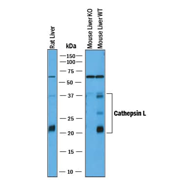
Detection of Mouse and Rat Cathepsin L by Western Blot.
Western blot shows lysates of rat liver tissue, mouse liver tissue (wild type), and mouse liver tissue (knock out). PVDF membrane was probed with 1 µg/mL of Goat Anti-Mouse Cathepsin L Antigen Affinity-purified Polyclonal Antibody (Catalog # AF1515) followed by HRP-conjugated Anti-Goat IgG Secondary Antibody (HAF017). Specific bands were detected for Cathepsin L at approximately 22-38 kDa (as indicated). This experiment was conducted under reducing conditions and using Immunoblot Buffer Group 1.Cathepsin L in Mouse Ovary.
Cathepsin L was detected in perfusion fixed frozen sections of mouse ovary using 15 µg/mL Goat Anti-Mouse Cathepsin L Antigen Affinity-purified Polyclonal Antibody (Catalog # AF1515) overnight at 4 °C. Tissue was stained with the Anti-Goat HRP-DAB Cell & Tissue Staining Kit (brown; CTS008) and counterstained with hematoxylin (blue). View our protocol for Chromogenic IHC Staining of Frozen Tissue Sections.Detection of Mouse Cathepsin L by Simple WesternTM.
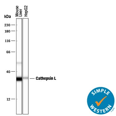
Simple Western lane view shows lysates of mouse liver tissue and HepG2 human hepatocellular carcinoma cell line, loaded at 0.2 mg/mL. Specific bands were detected for Cathepsin L at approximately 51 and 34 kDa (as indicated) using 10 µg/mL of Goat Anti-Mouse Cathepsin L Antigen Affinity-purified Polyclonal Antibody (Catalog # AF1515) followed by 1:50 dilution of HRP-conjugated Anti-Goat IgG Secondary Antibody (HAF109). This experiment was conducted under reducing conditions and using the 12-230 kDa separation system.Applications for Mouse/Rat Cathepsin L Antibody
Immunohistochemistry
Sample: Perfusion fixed frozen sections of mouse ovary and thymus
Simple Western
Sample: Mouse liver tissue and HepG2 human hepatocellular carcinoma cell line
Western Blot
Sample: Rat liver tissue, mouse liver tissue (wild type), and mouse liver tissue (knock out).
Reviewed Applications
Read 3 reviews rated 5 using AF1515 in the following applications:
Formulation, Preparation, and Storage
Purification
Reconstitution
Formulation
Shipping
Stability & Storage
- 12 months from date of receipt, -20 to -70 °C as supplied.
- 1 month, 2 to 8 °C under sterile conditions after reconstitution.
- 6 months, -20 to -70 °C under sterile conditions after reconstitution.
Background: Cathepsin L
Cathepsin L is a lysosomal cysteine protease expressed in most eukaryotic cells. Cathepsin L is known to hydrolyze a number of proteins, including the proform of urokinase-type plasminogen activator, which is activated by Cathepsin L cleavage (1). Cathepsin L has also been shown to proteolytically inactivate alpha1-antitrypsin and secretory leucoprotease inhibitor, two major protease inhibitors of the respiratory tract (2). These observations, combined with the demonstration of increased Cathepsin L activity in the epithelial lining fluid of the lungs of emphysema patients, have led to the suggestion that the enzyme may be involved in the progression of this disease. Cathepsin L has also been identified as a major excreted protein of transformed fibroblasts, indicating the enzyme could be involved in malignant tumor growth (3). In Cathepsin L-deficient mice, it appears to play a critical role in cardiac morphology and function, epidermal homeostasis, regulation of the hair cycle, and MHC class II-mediated antigen presentation in cortical epithelial cells of the thymus (4, 5). Mouse Cathepsin L is synthesized as a 334 amino acid precursor with a signal peptide (residues 1-17), a pro region (residues 18-113), and a mature chain (residues 114-334).
References
- Goretzki, L. et al. (1992) FEBS Lett. 297:112.
- Taggart, C.C. et al. (2001) J. Biol. Chem. 276:33345.
- Gottesman, M.M. and F. Cabral (1981) Biochemistry 20:1659.
- Stypmann, J. et al. (2002) Proc. Natl. Acad. Sci. USA 99: 6234.
- Reinheckel, T. et al. (2001) Biol. Chem. 382:735.
Alternate Names
Gene Symbol
UniProt
Additional Cathepsin L Products
Product Documents for Mouse/Rat Cathepsin L Antibody
Product Specific Notices for Mouse/Rat Cathepsin L Antibody
For research use only


