Mouse/Rat Complement Component C3d Antibody
R&D Systems, part of Bio-Techne | Catalog # AF2655


Key Product Details
Species Reactivity
Validated:
Mouse, Rat
Cited:
Human, Mouse, Rat, Transgenic Mouse
Applications
Validated:
Immunohistochemistry, Western Blot
Cited:
ELISA Capture, ELISA Development, Immunocytochemistry, Immunofluorescence, Immunohistochemistry, Immunohistochemistry-Frozen, Immunohistochemistry-Paraffin, Western Blot
Label
Unconjugated
Antibody Source
Polyclonal Goat IgG
Product Specifications
Immunogen
E. coli-derived recombinant mouse Complement Component C3d
His1002-Arg1303 (Cys1010Ser)
Accession # P01027
His1002-Arg1303 (Cys1010Ser)
Accession # P01027
Specificity
Detects mouse Complement Component C3d in direct ELISAs and Western blots. Detects mouse and rat Complement Component C3d in immunohistochemistry.
Clonality
Polyclonal
Host
Goat
Isotype
IgG
Scientific Data Images for Mouse/Rat Complement Component C3d Antibody
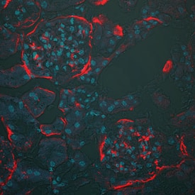
Complement Component C3d in Mouse Kidney.
Complement Component C3d was detected in perfusion fixed frozen sections of mouse kidney using Goat Anti-Mouse Complement Component C3d Antigen Affinity-purified Polyclonal Antibody (Catalog # AF2655) overnight at 4 °C. Tissue was stained using the NorthernLights™ 557-conjugated Anti-Goat IgG Secondary Antibody (red; Catalog # NL001) and counterstained with DAPI (blue). Specific staining was localized to basement membrane. View our protocol for Fluorescent IHC Staining of Frozen Tissue Sections.Complement Component C3d in Rat Kidney.
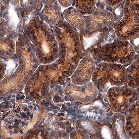
Complement Component C3d was detected in perfusion fixed paraffin-embedded sections of rat kidney using Goat Anti-Mouse Complement Component C3d Antigen Affinity-purified Polyclonal Antibody (Catalog # AF2655) at 3 µg/mL for 1 hour at room temperature followed by incubation with the Anti-Goat IgG VisUCyte™ HRP Polymer Antibody (Catalog # VC004). Before incubation with the primary antibody, tissue was subjected to heat-induced epitope retrieval using Antigen Retrieval Reagent-Basic (Catalog # CTS013). Tissue was stained using DAB (brown) and counterstained with hematoxylin (blue). Specific staining was localized to cytoplasm. View our protocol for IHC Staining with VisUCyte HRP Polymer Detection Reagents.Detection of Mouse Mouse/Rat Complement Component C3d Antibody by Immunohistochemistry
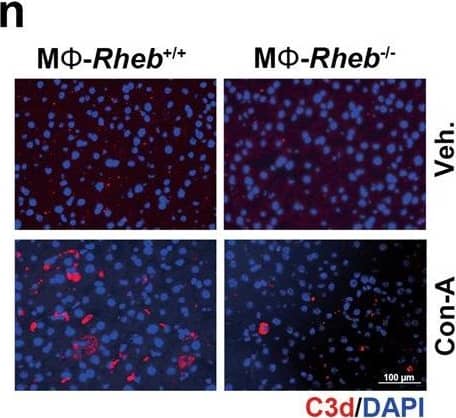
The activation of mTORC1 in KCs enhances complement alternative system.a Expression heatmap of genes of neutrophils chemotaxis or complement activation analyzed by RNA-seq from Tsc1+/+ and Tsc1-/- BMMs (n = 3 each). b Western blotting result was shown the expression of CFB protein in hepatic tissues from mice. c Left, representative co-immunofluorescent staining images for F4/80 with CFB. Scale bar = 50 μm. Right, quantitative determination of F4/80+ and CFB+ cells among groups as indicated, n = 3. d Western blotting assay showing the abundance for TSC1, CFB, and p-S6 in BMMs. e qRT-PCR analysis showing the CFB mRNA abundance in BMMs, n = 3. f Western blotting assay showing the abundance for TSC1, CFB, and p-S6 in KCs. g qRT-PCR analysis showing the CFB mRNA abundance in KCs, n = 3. h Representative immunofluorescent staining images for C3d. Scale bar = 50 μm. i Representative immunostaining images for C5b-9. Scale bar = 50 μm. j Western blotting assay showing the abundance for Rheb and CFB in BMMs. k Western blotting assay showing the abundance for Rheb and CFB in KCs. l Representative co-immunofluorescent staining images for F4/80 with CFB (white arrows). Scale bar = 100 μm. m Quantitative determination of F4/80+ and CFB+ cells among groups as indicated, n = 3. n Representative immunofluorescent staining images for C3d. Scale bar = 100 μm. o Representative immunostaining images for C5b-9 among groups as indicated. Scale bar = 100 μm. *p < 0.05. Image collected and cropped by CiteAb from the following publication (https://pubmed.ncbi.nlm.nih.gov/36494334), licensed under a CC-BY license. Not internally tested by R&D Systems.Applications for Mouse/Rat Complement Component C3d Antibody
Application
Recommended Usage
Immunohistochemistry
5-15 µg/mL
Sample: Perfusion fixed frozen sections of mouse kidney and perfusion fixed paraffin-embedded sections of rat kidney
Sample: Perfusion fixed frozen sections of mouse kidney and perfusion fixed paraffin-embedded sections of rat kidney
Western Blot
0.1 µg/mL
Sample: Recombinant Mouse Complement Component C3d
Sample: Recombinant Mouse Complement Component C3d
Reviewed Applications
Read 2 reviews rated 4 using AF2655 in the following applications:
Formulation, Preparation, and Storage
Purification
Antigen Affinity-purified
Reconstitution
Reconstitute at 0.2 mg/mL in sterile PBS. For liquid material, refer to CoA for concentration.
Formulation
Lyophilized from a 0.2 μm filtered solution in PBS with Trehalose. *Small pack size (SP) is supplied either lyophilized or as a 0.2 µm filtered solution in PBS.
Shipping
Lyophilized product is shipped at ambient temperature. Liquid small pack size (-SP) is shipped with polar packs. Upon receipt, store immediately at the temperature recommended below.
Stability & Storage
Use a manual defrost freezer and avoid repeated freeze-thaw cycles.
- 12 months from date of receipt, -20 to -70 °C as supplied.
- 1 month, 2 to 8 °C under sterile conditions after reconstitution.
- 6 months, -20 to -70 °C under sterile conditions after reconstitution.
Background: Complement Component C3d
Alternate Names
AHUS5;ARMD9;ASP;C3a;C3b;Complement C3;CPAMD1;HEL-S-62p
Entrez Gene IDs
718 (Human)
Gene Symbol
C3
UniProt
Additional Complement Component C3d Products
Product Documents for Mouse/Rat Complement Component C3d Antibody
Product Specific Notices for Mouse/Rat Complement Component C3d Antibody
For research use only
Loading...
Loading...
Loading...
Loading...
Loading...


