Mouse/Rat Neuropilin-2 Antibody
R&D Systems, part of Bio-Techne | Catalog # AF567


Key Product Details
Species Reactivity
Validated:
Cited:
Applications
Validated:
Cited:
Label
Antibody Source
Product Specifications
Immunogen
Gln23-Asp857 (Val809-Asp825 del)
Accession # O35276
Specificity
Clonality
Host
Isotype
Endotoxin Level
Scientific Data Images for Mouse/Rat Neuropilin-2 Antibody
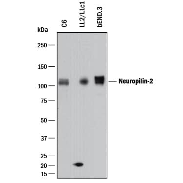
Detection of Mouse and Rat Neuropilin‑2 by Western Blot.
Western blot shows lysates of C6 rat glioma cell line, LL/2 mouse Lewis lung carcinoma cell line, and bEnd.3 mouse endothelioma cell line. PVDF membrane was probed with 0.5 µg/mL of Goat Anti-Mouse/Rat Neuropilin-2 Antigen Affinity-purified Polyclonal Antibody (Catalog # AF567) followed by HRP-conjugated Anti-Goat IgG Secondary Antibody (Catalog # HAF017). A specific band was detected for Neuropilin-2 at approximately 110 kDa (as indicated). This experiment was conducted under reducing conditions and using Immunoblot Buffer Group 1.Neuropilin‑2 in Rat Brain.
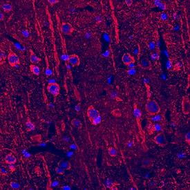
Neuropilin-2 was detected in perfusion fixed frozen sections of rat brain using Goat Anti-Mouse/Rat Neuropilin-2 Antigen Affinity-purified Polyclonal Antibody (Catalog # AF567) at 15 µg/mL overnight at 4 °C. Tissue was stained using the NorthernLights™ 557-conjugated Anti-Goat IgG Secondary Antibody (red; Catalog # NL001) and counterstained with DAPI (blue). Specific staining was localized to cytoplasm in neurons. View our protocol for Fluorescent IHC Staining of Frozen Tissue Sections.Detection of Mouse and Rat Neuropilin‑2 by Simple WesternTM.
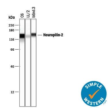
Simple Western lane view shows lysates of C6 rat glioma cell line, LL/2 mouse Lewis lung carcinoma cell line, and bEnd.3 mouse endothelioma cell line, loaded at 0.2 mg/mL. A specific band was detected for Neuropilin-2 at approximately 140 kDa (as indicated) using 5 µg/mL of Goat Anti-Mouse/Rat Neuropilin-2 Antigen Affinity-purified Polyclonal Antibody (Catalog # AF567) followed by 1:50 dilution of HRP-conjugated Anti-Goat IgG Secondary Antibody (Catalog # HAF109). This experiment was conducted under reducing conditions and using the 12-230 kDa separation system.Applications for Mouse/Rat Neuropilin-2 Antibody
Blockade of Receptor-ligand Interaction
Immunohistochemistry
Sample: Perfusion fixed frozen sections of rat brain
Simple Western
Sample: C6 rat glioma cell line, LL/2 mouse Lewis lung carcinoma cell line, and bEnd.3 mouse endothelioma cell line
Western Blot
Sample: C6 rat glioma cell line, LL/2 mouse Lewis lung carcinoma cell line, and bEnd.3 mouse endothelioma cell line
Reviewed Applications
Read 7 reviews rated 4.4 using AF567 in the following applications:
Formulation, Preparation, and Storage
Purification
Reconstitution
Formulation
Shipping
Stability & Storage
- 12 months from date of receipt, -20 to -70 °C as supplied.
- 1 month, 2 to 8 °C under sterile conditions after reconstitution.
- 6 months, -20 to -70 °C under sterile conditions after reconstitution.
Background: Neuropilin-2
Neuropilin-1 (Npn-1, previously known as Neuropilin) and Npn-2 (previously known as Npn-1-related molecule) are type I transmembrane proteins that bind members of the class III secreted semaphorin subfamily which are implicated in repulsive axon guidance. The extracellular domain of these proteins is composed of two N-terminal CUB (complement-binding) domains (domains a1 and a2), two domains with homology to coagulation factors V and VIII (domains b1 and b2) and a MAM (meprin) domain (domain c). In the absence of ligands, neuropilins can form homo- and hetero-oligomers via homophilic interactions of their MAM domains. At the amino acid sequence level, Npn-1and Npn-2 share 44% identity. Npn-1 and Npn-2 show different binding specificities for different members of the semaphorin family. The expression patterns of Npn-1 and Npn-2 in developing neurons of the central and peripheral nervous systems are largely, though not completely nonoverlapping. Npn‑1 and Npn-2 are also expressed by endothelial and tumor cells and have been shown to be isoform-specific receptors for VEGF165. Npn‑1 was also reported to bind PlGF-2 and the VEGF-like protein from of virus NZ2.
References
- Fujisawa, H. and T. Kitsukawa (1998) Curr. Opin. Neurobiol. 8:587.
- Neufeld, G. et al. (1999) FASEB J. 13:9.
- Poltorak, Z. et al. (2000) J. Biol. Chem. 275:18040.
Alternate Names
Gene Symbol
UniProt
Additional Neuropilin-2 Products
Product Documents for Mouse/Rat Neuropilin-2 Antibody
Product Specific Notices for Mouse/Rat Neuropilin-2 Antibody
This product or the use of this product is covered by U.S. Patents owned by The Regents of the University of California. This product is for research use only and is not to be used for commercial purposes. Use of this product to produce products for sale or for diagnostic, therapeutic or drug discovery purposes is prohibited. In order to obtain a license to use this product for such purposes, contact The Regents of the University of California.
U.S. Patent # 6,054,293, 6,623,738, and other U.S. and international patents pending.
For research use only

