Mouse RGM-B Antibody
R&D Systems, part of Bio-Techne | Catalog # AF3597


Key Product Details
Species Reactivity
Validated:
Cited:
Applications
Validated:
Cited:
Label
Antibody Source
Product Specifications
Immunogen
Gly49-Ser414
Accession # Q7TQ33
Specificity
Clonality
Host
Isotype
Scientific Data Images for Mouse RGM-B Antibody
RGM-B in Mouse Brain.
RGM-B was detected in perfusion fixed frozen sections of mouse brain (trigeminal ganglia) using 1.7 µg/mL Sheep Anti-Mouse RGM-B Antigen Affinity-purified Polyclonal Antibody (Catalog # AF3597) overnight at 4 °C. Tissue was stained with the Anti-Sheep HRP-DAB Cell & Tissue Staining Kit (brown; Catalog # CTS019) and counterstained with hematoxylin (blue). View our protocol for Chromogenic IHC Staining of Frozen Tissue Sections.Detection of RGM-B by Immunohistochemistry
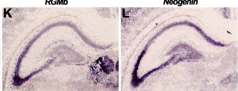
Subregion-specific expression of RGMs and Neogenin in the hippocampus.In situ hybridization (A–C, J–L) and immunohistochemistry (D–I, M–O) on coronal mouse brain sections at E16.5 (A–I) and P5 (J–O). Sections in D–I and M–O are counterstained in blue with fluorescent Nissl. (A–F) RGMa mRNA and protein are expressed in the ventricular zone (VZ), dentate gyrus (DG) and cornu ammonis (CA) region. Strong expression of RGMb mRNA and protein is detected in the pial surface lining the hippocampal fissure (HF). Neogenin transcripts and protein are widely expressed in the developing hippocampus (Hip). (G–I) Immunostaining with isotype matched controls. (J–L) In situ hybridization at P5 shows strong but differential expression patterns of RGMa, RGMb and Neogenin in the CA pyramidal cell layers (Pyr). In addition, strong expression of Neogenin is detected in the granular layer (GC) of the DG. (M–O) Immunohistochemistry reveals expression of RGMa and weak expression of RGMb in the stratum lacunosum moleculare (SLM) and fimbria (FIM). Neogenin strongly labels different hippocampal layers. CX, cortex; Hb, habenula; HC, hippocampal commissure; PO, polymorph layer; SO, stratum oriens; SR, stratum radiatum; Th, thalamus. Scale bar A–C: 400 µm, D–F: 300 µm, G–I: 300 µm, J–L: 500 µm and M–O: 400 µm. Image collected and cropped by CiteAb from the following open publication (https://pubmed.ncbi.nlm.nih.gov/23457482), licensed under a CC-BY license. Not internally tested by R&D Systems.Detection of RGM-B by Immunohistochemistry
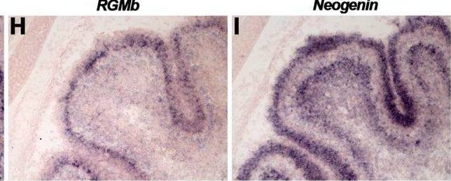
Differential expression of RGMs and Neogenin in the cerebellum.In situ hybridization (A–C, G–I, M–O) and immunohistochemistry (D–F, J–L, P–R) on coronal mouse brain sections at E16.5 (A–F), P5 (G–L) and in the adult (M–R). (A–I, M–O) In situ hybridization and immunohistochemistry reveals strong and broad expression of RGMa, RGMb and Neogenin in the cerebellum (CB). (J–L) Immunostaining shows expression of RGMa and Neogenin in all cerebellar layers at P5. RGMb is expressed in the internal granular layer (IGL), Purkinje cell layer (PCL) and external granular layer (EGL). (P–R) In the adult, RGMa, RGMb and Neogenin protein are expressed in Purkinje cells (PCs) and axons in the granular cell layer (GCL), PCL and molecular layer (ML). Neogenin strongly labels PC dendrites in the ML. DCN, deep cerebellar nuclei; WM, white matter; VZ, ventricular zone. Scale bar A–C: 300 µm, D–F: 250 µm, G–I: 150 µm, J–L: 150 µm, M–O: 100 µm and P–R: 50 µm. Image collected and cropped by CiteAb from the following open publication (https://pubmed.ncbi.nlm.nih.gov/23457482), licensed under a CC-BY license. Not internally tested by R&D Systems.Applications for Mouse RGM-B Antibody
CyTOF-ready
Flow Cytometry
Sample: Neuro-2A mouse neuroblastoma cell line
Immunohistochemistry
Sample: Perfusion fixed frozen sections of mouse brain (trigeminal ganglia)
Western Blot
Sample: Recombinant Mouse RGM-B (Catalog # 3597-RG)
Formulation, Preparation, and Storage
Purification
Reconstitution
Formulation
Shipping
Stability & Storage
- 12 months from date of receipt, -20 to -70 °C as supplied.
- 1 month, 2 to 8 °C under sterile conditions after reconstitution.
- 6 months, -20 to -70 °C under sterile conditions after reconstitution.
Background: RGM-B
RGM-B, also known as DRAGON, is a 40 kDa member of the repulsive guidance molecule (RGM) family of GPI-linked neuronal and muscle membrane proteins (1, 2). It is synthesized as a preproprotein that consists of a 48 amino acid (aa) signal sequence, a 367 aa mature region, and a 21 aa C-terminal prosegment (3). RGM-B contains two potential N-linked glycosylation sites and an abbreviated von Willebrand factor domain. Potential proteolytic cleavage within the VWF domain is supported by R&D Systems’ in house data (4). Within the region following the VWF domain, mouse RGM-B shares 49% and 43% aa sequence identity with RGM-A and RGM-C, respectively. It shares 90%, 79%, 92%, and 93% aa sequence identity with bovine, chicken, human, and rhesus macaque RGM-B, respectively. RGM-B is expressed in the developing and adult nervous system, particularly in the dorsal root ganglia and mantle layer of the spinal cord (3-5). In mouse, it shows a complementary, non-overlapping distribution with RGM-A (2-5). RGM-B is also expressed in fetal and adult enteric ganglia and in postnatal intestinal epithelium (6). RGM-B expression has been detected in neuronal cell bodies and proximal axonal segments (4) but is also present on the cell surface, where it interacts homophilically and mediates neuronal adhesion (3). RGM-B additionally functions as a BMP coreceptor. It directly binds BMP-2 and -4 but not other TGF-beta family proteins (7). RGM-B associates with BMP type I (ALK-2, -3, -6) and type II (Activin RIIA, Activin RIIB) receptors and enhances BMP signaling (7).
References
- Monnier, P.P. et al. (2002) Nature 419:392.
- Schmidtmer, J. and D. Engelkamp (2004) Gene Exp. Patterns 4:105.
- Samad, T.A. et al. (2004) J. Neurosci. 24:2027.
- Niederkofler, V. et al. (2004) J. Neurosci. 24:808.
- Oldekamp, J. et al. (2004) Gene Exp. Patterns 4:283.
- Metzger, M. et al. (2005) Dev. Dyn. 234:169.
- Samad, T.A. et al. (2005) J. Biol. Chem. 280:14122.
Long Name
Alternate Names
Gene Symbol
UniProt
Additional RGM-B Products
Product Documents for Mouse RGM-B Antibody
Product Specific Notices for Mouse RGM-B Antibody
For research use only


