Mouse Sca-1/Ly6 Antibody
R&D Systems, part of Bio-Techne | Catalog # AF1226


Key Product Details
Species Reactivity
Validated:
Cited:
Applications
Validated:
Cited:
Label
Antibody Source
Product Specifications
Immunogen
Leu27-Gly119
Accession # CAA28351
Specificity
Clonality
Host
Isotype
Scientific Data Images for Mouse Sca-1/Ly6 Antibody
Sca-1/Ly6 in Mouse Splenocytes.
Sca-1/Ly6 was detected in immersion fixed mouse splenocytes using 10 µg/mL Goat Anti-Mouse Sca-1/Ly6 Antigen Affinity-purified Polyclonal Antibody (Catalog # AF1226) for 3 hours at room temperature. Cells were stained with the NorthernLights™ 557-conjugated Anti-Goat IgG Secondary Antibody (red; Catalog # NL001) and counterstained with DAPI (blue). View our protocol for Fluorescent ICC Staining of Non-adherent Cells.Detection of Mouse Sca-1/Ly6 by Western Blot
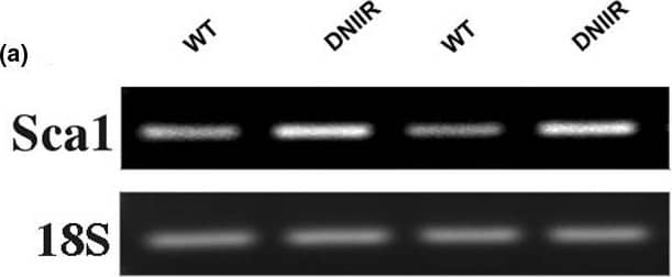
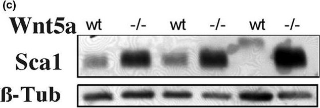
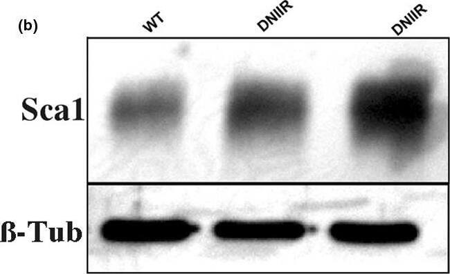
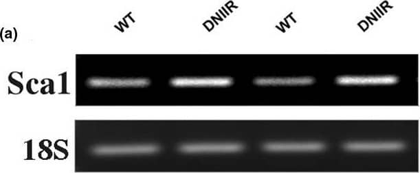
Sca1, a marker of mammary stem/progenitor cells, is increased in DNIIR and Wnt5a-/- glands. mRNA and protein were extracted from several wild type (WT) and MT-DNIIR (DNIIR) mice. (a) Sca1 mRNA levels were determined by semi-quantitative RT-PCR and (b) protein levels were determined by western blot. Sca1 levels were increased in DNIIR glands relative to controls. (c) Sca1 protein levels were determined in WT and Wnt5a null (-/-) glands using western blot analysis. Sca1 levels were increased in Wnt5a-null tissue relative to controls. 18S and beta-Tubulin ( beta-Tub) were used as normalisation controls. DNIIR = dominant-negative transforming growth factor-beta type II receptor; RT-PCR = reverse transcripase polymerase chain reaction; Wnt = Wingless-related protein family. Image collected and cropped by CiteAb from the following open publication (https://pubmed.ncbi.nlm.nih.gov/19344510), licensed under a CC-BY license. Not internally tested by R&D Systems.Detection of Mouse Sca-1/Ly6 by Western Blot
Sca1, a marker of mammary stem/progenitor cells, is increased in DNIIR and Wnt5a-/- glands. mRNA and protein were extracted from several wild type (WT) and MT-DNIIR (DNIIR) mice. (a) Sca1 mRNA levels were determined by semi-quantitative RT-PCR and (b) protein levels were determined by western blot. Sca1 levels were increased in DNIIR glands relative to controls. (c) Sca1 protein levels were determined in WT and Wnt5a null (-/-) glands using western blot analysis. Sca1 levels were increased in Wnt5a-null tissue relative to controls. 18S and beta-Tubulin ( beta-Tub) were used as normalisation controls. DNIIR = dominant-negative transforming growth factor-beta type II receptor; RT-PCR = reverse transcripase polymerase chain reaction; Wnt = Wingless-related protein family. Image collected and cropped by CiteAb from the following open publication (https://pubmed.ncbi.nlm.nih.gov/19344510), licensed under a CC-BY license. Not internally tested by R&D Systems.Applications for Mouse Sca-1/Ly6 Antibody
CyTOF-ready
Flow Cytometry
Sample: Mouse splenocytes
Immunocytochemistry
Sample: Immersion fixed mouse splenocytes
Western Blot
Sample: Recombinant Mouse Sca-1/Ly6
Reviewed Applications
Read 1 review rated 5 using AF1226 in the following applications:
Formulation, Preparation, and Storage
Purification
Reconstitution
Formulation
Shipping
Stability & Storage
- 12 months from date of receipt, -20 to -70 °C as supplied.
- 1 month, 2 to 8 °C under sterile conditions after reconstitution.
- 6 months, -20 to -70 °C under sterile conditions after reconstitution.
Background: Sca-1/Ly6
This antibody reacts with Sca-1, an 18 kDa phosphatidylinositol-anchored protein that is a member of the lymphocyte antigen 6 (Ly-6) family (1). Sca-1 is encoded by the strain-specific Ly-6 E/A allelic gene. Its expression on multipotent hematopoietic stem cells (HSC) has been used as a marker of HSC in mice of both Ly‑6 haplotypes (2, 3). This antibody is frequently used in combination with lineage depletion antibodies to identify and isolate murine HSC. Sca-1-positive HSC can be found in the adult bone marrow, fetal liver and mobilized peripheral blood and spleen in the adult animal (2‑7). However, Sca-1 has also been discovered in several non‑hematopoietic tissues (1) and can be used to enrich progenitor cell populations other than HSC (8). It is suggested that Sca-1 could be involved in regulating both B and T cell activation (9‑12).
References
- Van de Rijn, M. et al. (1989) Proc. Natl.Acad. Sci. USA 86:4634.
- Spangrude, G.I. et al. (1988) Science 241:58.
- Spangrude, G.I. et al. (1993) Blood 82:3327.
- Morrison, S.J. et al. (1995) Proc. Natl. Acad. Sci. USA 92:10302.
- Kawamoto, H. et al. (1997) Int. Immunol. 9:1011.
- Yamamoto, Y. et al. (1996) Blood 88:445.
- Morrison, S.J. et al. (1997) Proc. Natl. Acad. Sci. USA 94:1908.
- Welm, B.E. et al. (2002) Dev. Biol. 245:42.
- Codias, E.K. et al. (1990) J. Immunol. 144:2197.
- Malek, T.R. et al. (1986) J. Exp. Med. 164:709 .
- Codias, E.K. et al. (1990) J. Immunol. 145:1407.
- Flood, P.M. et al. (1990) J. Exp. Med. 172:115.
Long Name
Alternate Names
Entrez Gene IDs
Gene Symbol
UniProt
Additional Sca-1/Ly6 Products
Product Documents for Mouse Sca-1/Ly6 Antibody
Product Specific Notices for Mouse Sca-1/Ly6 Antibody
For research use only
