Mouse Serpin A1c/ alpha1-Antitrypsin Antibody
R&D Systems, part of Bio-Techne | Catalog # AF2979

Key Product Details
Species Reactivity
Validated:
Cited:
Applications
Validated:
Cited:
Label
Antibody Source
Product Specifications
Immunogen
Glu25-Lys413
Accession # NP_033271
Specificity
Clonality
Host
Isotype
Scientific Data Images for Mouse Serpin A1c/ alpha1-Antitrypsin Antibody
Detection of Mouse Serpin A1c/ alpha1‑Antitrypsin by Western Blot.
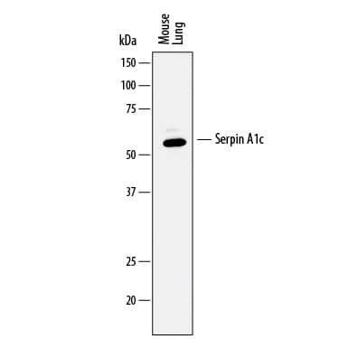
Western blot shows lysates of mouse lung tissue. PVDF membrane was probed with 0.5 µg/mL of Goat Anti-Mouse Serpin A1c/a1-Antitrypsin Antigen Affinity-purified Polyclonal Antibody (Catalog # AF2979) followed by HRP-conjugated Anti-Goat IgG Secondary Antibody (Catalog # HAF017). A specific band was detected for Serpin A1c/a1-Antitrypsin at approximately 55 kDa (as indicated). This experiment was conducted under reducing conditions and using Immunoblot Buffer Group 1.Detection of Mouse Serpin A1c/ alpha1‑Antitrypsin by Simple WesternTM.
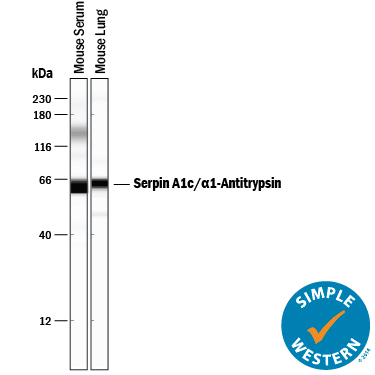
Simple Western lane view shows mouse serum and lysates of mouse lung tissue, loaded at 0.2 mg/mL. A specific band was detected for Serpin A1c/a1-Antitrypsin at approximately 62 kDa (as indicated) using 5 µg/mL of Goat Anti-Mouse Serpin A1c/a1-Antitrypsin Antigen Affinity-purified Polyclonal Antibody (Catalog # AF2979) followed by 1:50 dilution of HRP-conjugated Anti-Goat IgG Secondary Antibody (Catalog # HAF109). This experiment was conducted under reducing conditions and using the 12-230 kDa separation system.Detection of Monkey Serpin A1c/alpha 1-Antitrypsin by Immunocytochemistry/Immunofluorescence
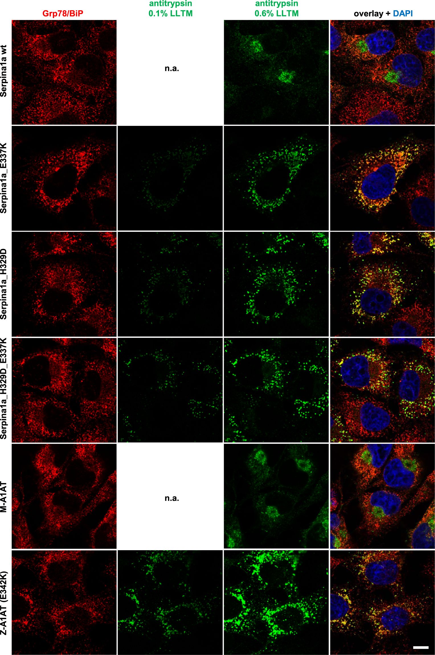
Serpina1a mutants imitate intracellular distribution of human Z-A1AT. Confocal laser immunofluorescence analysis of COS-7 cells expressing wild type (wt; top row), E337K, H329D or H329D_E337K double mutant Serpina1a (second to fourth row), compared to cells overexpressing normal human M-A1AT or E342K Z-A1AT (fifth and sixth row). Mouse Serpina1a and human A1AT were stained with Alexa Fluor 568 secondary antibody (green) and exposed to 0.6% laser light transmission (LLTM). Mutant-expressing cells were additionally exposed to 0.1% LLTM, as the very strong signal resulted in over-saturation at 0.6%. ER-marker Grp78/BiP was stained with Alexa Fluor 647 secondary antibody (red) and cell nucleus was stained using DAPI (blue). Scale bar: 10 µm. Image collected and cropped by CiteAb from the following publication (https://pubmed.ncbi.nlm.nih.gov/31097772), licensed under a CC-BY license. Not internally tested by R&D Systems.Applications for Mouse Serpin A1c/ alpha1-Antitrypsin Antibody
Simple Western
Sample: Mouse serum and mouse lung tissue
Western Blot
Sample: Mouse lung tissue
Formulation, Preparation, and Storage
Purification
Reconstitution
Formulation
Shipping
Stability & Storage
- 12 months from date of receipt, -20 to -70 °C as supplied.
- 1 month, 2 to 8 °C under sterile conditions after reconstitution.
- 6 months, -20 to -70 °C under sterile conditions after reconstitution.
Background: Serpin A1c/alpha 1-Antitrypsin
SerpinA1c (serine proteinase inhibitor-clade A 1c; also alpha-1 protease inhibitor 6 and alpha1 antitrypsin 1-3) is a secreted, 50-55 kDa glycoprotein member of the clade A-subfamily, serpin superfamily of protease inhibitors. There are multiple sources for serpinA1. The circulating form of serpinA1 is expressed by hepatocytes, while local production occurs in the bone marrow by osteoblasts, PMNs, T cells and B cells. SerpinA1 is a naturally occurring serine protease inhibitor. Its principal activity seems to be that of neutralizing neutrophil elastase, an activity that protects the elasticity of the lung during inflammation. It is also posited to play a role in bone marrow progenitor mobilization. Here, it promotes HPC proliferation by blocking cytokine degradation, and interferes with HPC migration by blocking protease cleavage of engaged cell adhesion molecules. Mature mouse serpinA1c is 389 amino acids (aa) in length (aa 25-413) (GenBank#:NP_033271) and contains one active enzymatic site (aa 67-410). Unlike human and rat, the mouse genus contains anywhere from one to seven serpinA1genes. Mus caroli (an Asian species) possesses one gene, Mus saxicola (a south asia species) possesses four genes, and Mus musculus possesses six or more distinct genes. Within Mus musculus, various strains have variable numbers of active genes. While highly homologous, the protein sequences are not identical, and differ most importantly in the reactive center loop region (aa 374-386). The protein referenced here is equivalent to alpha1-PI-3 (Borriello, F & K.S. Krauter [1991] Proc. Natl. Acad. Sci. USA 88:9417). This gene has one isoform with minimal scattered substitutions and shows more that 98% aa sequence identity (GenBank #:Q00896). Over aa 25-413, serpinA1c/ alpha1-IP-3 shares 97% and 95% aa sequence identity with serpinA1a/ alpha1-PI-1 and serpinA1b/ alpha1-IP-2, respectively. It also shares 77% and 64% aa sequence identity with rat and human serpinA1, respectively.
Alternate Names
Gene Symbol
UniProt
Additional Serpin A1c/alpha 1-Antitrypsin Products
Product Documents for Mouse Serpin A1c/ alpha1-Antitrypsin Antibody
Product Specific Notices for Mouse Serpin A1c/ alpha1-Antitrypsin Antibody
For research use only


