Mouse Siglec-F Biotinylated Antibody
R&D Systems, part of Bio-Techne | Catalog # BAF1706


Key Product Details
Species Reactivity
Validated:
Cited:
Applications
Validated:
Cited:
Label
Antibody Source
Product Specifications
Immunogen
Asp18-Thr437
Accession # Q920G3
Specificity
Clonality
Host
Isotype
Scientific Data Images for Mouse Siglec-F Biotinylated Antibody
Detection of Mouse Siglec-F by Immunocytochemistry/ Immunofluorescence
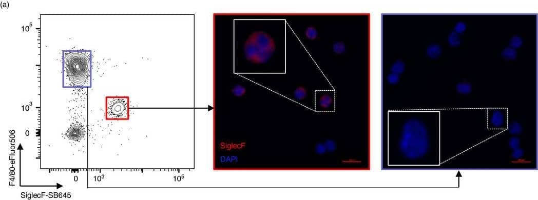
Wild house mice harbour a large pool of eosinophils in spleen and bone marrow. Spleens, peritoneal exudate cells (PEC) and bone marrow (BM) were collected from wild house mice from the Isle of May between September and December 2019, as well as from naïve C57BL/6 mice. (a) PEC cells from wild mice FACS sorted on either Siglec‐F+F4/80int or Siglec‐F−F4/80hi were cytospun and stained with DAPI (blue) and anti‐Siglec‐F antibody (red); n = 1. (b) ImageStream analysis of wild mouse splenic Siglec‐F+ cells, representative of 5 mice. (c) Proportion of Siglec‐F+F4/80int eosinophils and Siglec‐F−F4/80−Ly6G+ neutrophils among the myeloid cell population in laboratory and wild mice. Box plots show median with interquartile range. Laboratory versus wild eosinophil proportions were analysed using a two‐tailed Mann–Whitney U‐test; n(Lab) = 16 (Spleen, PEC), 10 (BM); n(Wild) = 25 (Spleen, PEC), 10 (BM) Image collected and cropped by CiteAb from the following open publication (https://pubmed.ncbi.nlm.nih.gov/34486729), licensed under a CC-BY license. Not internally tested by R&D Systems.Detection of Mouse Siglec-F by Immunocytochemistry/ Immunofluorescence
Wild house mice harbour a large pool of eosinophils in spleen and bone marrow. Spleens, peritoneal exudate cells (PEC) and bone marrow (BM) were collected from wild house mice from the Isle of May between September and December 2019, as well as from naïve C57BL/6 mice. (a) PEC cells from wild mice FACS sorted on either Siglec‐F+F4/80int or Siglec‐F−F4/80hi were cytospun and stained with DAPI (blue) and anti‐Siglec‐F antibody (red); n = 1. (b) ImageStream analysis of wild mouse splenic Siglec‐F+ cells, representative of 5 mice. (c) Proportion of Siglec‐F+F4/80int eosinophils and Siglec‐F−F4/80−Ly6G+ neutrophils among the myeloid cell population in laboratory and wild mice. Box plots show median with interquartile range. Laboratory versus wild eosinophil proportions were analysed using a two‐tailed Mann–Whitney U‐test; n(Lab) = 16 (Spleen, PEC), 10 (BM); n(Wild) = 25 (Spleen, PEC), 10 (BM) Image collected and cropped by CiteAb from the following open publication (https://pubmed.ncbi.nlm.nih.gov/34486729), licensed under a CC-BY license. Not internally tested by R&D Systems.Applications for Mouse Siglec-F Biotinylated Antibody
Western Blot
Sample: Recombinant Mouse Siglec-F Fc Chimera (Catalog # 1706-SF)
Formulation, Preparation, and Storage
Purification
Reconstitution
Formulation
Shipping
Stability & Storage
- 12 months from date of receipt, -20 to -70 °C as supplied.
- 1 month, 2 to 8 °C under sterile conditions after reconstitution.
- 6 months, -20 to -70 °C under sterile conditions after reconstitution.
Background: Siglec-F
Siglecs (1) (sialic acid binding Ig-like lectins) are I-type (Ig-type) lectins (2) belonging to the Ig superfamily. They are characterized by an N-terminal Ig-like V-type domain which mediates sialic acid binding (3), followed by varying numbers of Ig-like C2-type domains (1, 4). Eleven human Siglecs have been cloned and characterized (1, 4). They are sialoadhesin/CD169/Siglec-1, CD22/Siglec-2, CD33/Siglec-3, Myelin-Associated Glycoprotein (MAG/Siglec-4a) and Siglec 5 to 11 (4‑6). To date, no Siglec has been shown to recognized any cell surface ligand other than sialic acids, suggesting that interactions with glycans containing this carbohydrate are important in mediating the biological functions of Siglecs. Siglec 5 to 11 share a high degree of sequence similarity with CD33/Siglec-3 both in their extracellular and intracellular regions. They are collectively referred to as CD33-related Siglecs. One remarkable feature of the CD33-related Siglecs is their differential expression pattern within the hematopoietic system (4, 5). This fact, together with the presence of two conserved immunoreceptor tyrosine-based inhibition motifs (ITIMs) in their cytoplasmic tails, suggests that CD33-related Siglecs are involved in the regulation of cellular activation within the immune system.
Mouse Siglec-F cDNA encodes a 569 amino acid polypeptide with a hydrophobic signal peptide, an N-terminal Ig-like V-type domain, three Ig-like C2-type domains, a transmembrane region and a cytoplasmic tail (7). The expression of Siglec-F is restricted to the cells of myelomonocytic lineage. Mouse Siglec-F is likely an ortholog of human Siglec-5. Unlike many human CD33-related Siglecs, which show similar binding to both alpha2,3- and alpha2,6-linked sialic acids, mouse Siglec-F preferentially recognize alpha2,3-linked sialic acid.
References
- Crocker, P.R. et al. (1998) Glycobiology 8:v.
- Powell, L.D. et al. (1995) J. Biol. Chem. 270:14243.
- May, A.R. et al. (1998) Mol. Cell 1998. 1:719.
- Crocker, P.R. and A. Varki (2001) Trends Immunol. 22:337.
- Crocker, P.R. et al. (2001) Immunology 103:137.
- Angata, T. et al. (2002) J. Biol Chem. 277:24466.
- Angata, T. et al. (2001) J. Biol Chem. 276:45128.
Long Name
Alternate Names
Entrez Gene IDs
Gene Symbol
UniProt
Additional Siglec-F Products
Product Documents for Mouse Siglec-F Biotinylated Antibody
Product Specific Notices for Mouse Siglec-F Biotinylated Antibody
For research use only
