Mouse Sortilin Antibody
R&D Systems, part of Bio-Techne | Catalog # AF2934


Key Product Details
Species Reactivity
Validated:
Cited:
Applications
Validated:
Cited:
Label
Antibody Source
Product Specifications
Immunogen
Gly76-Asn753
Accession # Q6PHU5
Specificity
Clonality
Host
Isotype
Endotoxin Level
Scientific Data Images for Mouse Sortilin Antibody
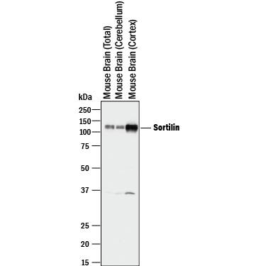
Detection of Mouse Sortilin by Western Blot.
Western blot shows lysates of mouse brain (total) tissue, mouse brain (cerebellum) tissue, and mouse brain (cortex) tissue. PVDF membrane was probed with 1 µg/mL of Goat Anti-Mouse Sortilin Antigen Affinity-purified Polyclonal Antibody (Catalog # AF2934) followed by HRP-conjugated Anti-Goat IgG Secondary Antibody (Catalog # HAF017). A specific band was detected for Sortilin at approximately 110 kDa (as indicated). This experiment was conducted under reducing conditions and using Immunoblot Buffer Group 1.Sortilin in Mouse Brain.
Sortilin was detected in perfusion fixed frozen sections of mouse brain (rostral ventral medulla) using Goat Anti-Mouse Sortilin Antigen Affinity-purified Polyclonal Antibody (Catalog # AF2934) at 1.7 µg/mL overnight at 4 °C. Tissue was stained using the Anti-Goat HRP-DAB Cell & Tissue Staining Kit (brown; Catalog # CTS008) and counterstained with hematoxylin (blue). View our protocol for Chromogenic IHC Staining of Frozen Tissue Sections.Detection of Mouse Sortilin by Simple WesternTM.
Simple Western lane view shows lysates of mouse brain (cortex) tissue , loaded at 0.2 mg/mL. A specific band was detected for Sortilin at approximately 122 kDa (as indicated) using 10 µg/mL of Goat Anti-Mouse Sortilin Antigen Affinity-purified Polyclonal Antibody (Catalog # AF2934) followed by 1:50 dilution of HRP-conjugated Anti-Goat IgG Secondary Antibody (Catalog # HAF109). This experiment was conducted under reducing conditions and using the 12-230 kDa separation system.Applications for Mouse Sortilin Antibody
Blockade of Receptor-ligand Interaction
Immunohistochemistry
Sample: Perfusion fixed frozen sections of mouse brain (rostral ventral medulla)
Simple Western
Sample: Mouse brain (cortex) tissue
Western Blot
Sample: Mouse brain (total) tissue, mouse brain (cerebellum) tissue, and mouse brain (cortex) tissue
Reviewed Applications
Read 8 reviews rated 5 using AF2934 in the following applications:
Formulation, Preparation, and Storage
Purification
Reconstitution
Formulation
Shipping
Stability & Storage
- 12 months from date of receipt, -20 to -70 °C as supplied.
- 1 month, 2 to 8 °C under sterile conditions after reconstitution.
- 6 months, -20 to -70 °C under sterile conditions after reconstitution.
Background: Sortilin
Sortilin (neurotensin receptor 3, glycoprotein 95) is a 95 kDa Type I transmembrane monomeric glycoprotein that is one of five known members of the mammalian vacuolar protein sorting 10p domain (Vps10p-D) family of sorting receptors (1, 2). Mouse preprosortilin is processed by signal sequence cleavage followed by propeptide cleavage at a furin recognition site. The cationic propeptide exhibits pH-dependent high affinity binding that blocks the Sortilin ligand binding site both pre‑ and post-cleavage (3). The extracellular/luminal sequence comprises the Vps10p domain, including 10 conserved cysteines (10CC) essential for ligand binding (2). The cytoplasmic domain sorting motifs confer all trafficking during synthesis, targeting to lysosomes, endocytosis and Golgi-endosome transport; as little as 10% may be found on the cell surface (4). Mature mouse Sortilin shares 98% amino acid (aa) identity with rat, and 91% aa identity with human and canine sortilin. During murine development, sortilin is mainly expressed in the nervous system (5), where it is a receptor for neuropeptides including neurotensin, nerve growth factor (NGF) and brain-derived neurotrophic factor (BDNF) (6‑9). ProNGF (or the NGF propeptide alone) binds sortilin with a much higher affinity (KD ~5-8 nM) than does mature NGF (KD ~90 nM). The complex of sortilin, pro‑NGF and the receptor p75ntr results in endocytosis of proNGF and induction of apoptosis (7). Similar results have been obtained with proBDNF and BDNF (8‑9). Sortilin is expressed in other tissues including testis, skeletal muscle and fat (1, 10). It is essential and sufficient for biogenesis of Glut4 storage vesicles necessary for insulin responsiveness in adipocytes (10). Sortilin also binds lipoprotein lipase (11), apoE (2) and RAP (1, 11). Binding is competitive, indicating that although unrelated, targets likely bind the same site.
References
- Petersen, C.M. et al. (1997) J. Biol. Chem. 272:3599.
- Westergaard, U.B. et al. (2004) J. Biol. Chem. 279:50221.
- Petersen, C.M. et al. (1998) EMBO J. 18:595.
- Nielsen, M.S. et al. (2001) EMBO J. 20:2180.
- Hermans-Borgmeyer, I. et al. (1999) Mol. Brain Res. 65:216.
- Mazella, J. et al. (1998) J. Biol. Chem. 273:26273.
- Nykjaer, A et al. (2004) Nature 427:843.
- Teng, H.K. et al. (2005) J. Neurosci. 25:5455.
- Chen, Z.-Y. et al. (2004) J. Neurosci. 25:6156.
- Shi, J and K.V. Kandror (2005) Dev. Cell 9:99.
- Nielsen, M.S. et al. (1999) J. Biol. Chem. 274:8832.
Alternate Names
Gene Symbol
UniProt
Additional Sortilin Products
Product Documents for Mouse Sortilin Antibody
Product Specific Notices for Mouse Sortilin Antibody
For research use only

