Mouse TCF-3/E2A Antibody

R&D Systems, part of Bio-Techne | Catalog # MAB7650

R&D Systems, part of Bio-Techne
Catalog #
Availability
Size / Price
Qty
Loading...
MAB7650
MAB7650-SP

Key Product Details

Species Reactivity

Validated:

Mouse

Cited:

Mouse

Applications

Validated:

Immunohistochemistry, Western Blot

Cited:

Western Blot

Label

Unconjugated

Antibody Source

Monoclonal Rat IgG2A Clone # 826927

Product Specifications

Immunogen

E. coli-derived recombinant mouse TCF‑3/E2A
Asn33-Arg159
Accession # P15806

Specificity

Detects mouse TCF‑3/E2A in ELISAs. In direct ELISAs, no cross-reactivity with recombinant human TCF-3 is observed.

Clonality

Monoclonal

Host

Rat

Isotype

IgG2A

Scientific Data Images for Mouse TCF-3/E2A Antibody

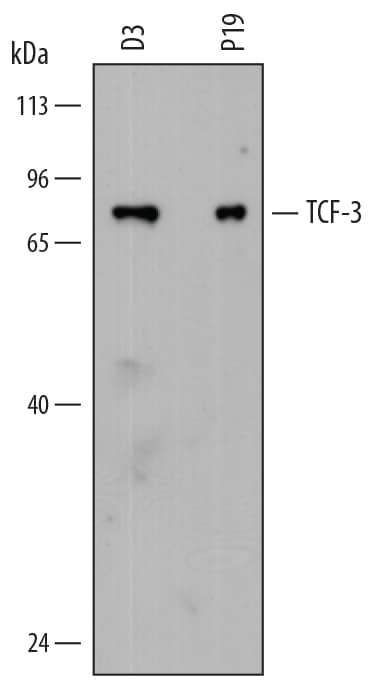
Detection of Mouse TCF-3/E2A antibody by Western Blot.

Detection of Mouse TCF‑3/E2A by Western Blot.

Western blot shows lysates of D3 mouse embryonic stem cell line and P19 mouse embryonal carcinoma cell line. PVDF membrane was probed with 2 µg/mL of Rat Anti-Mouse TCF-3/E2A Monoclonal Antibody (Catalog # MAB7650) followed by HRP-conjugated Anti-Rat IgG Secondary Antibody (Catalog # HAF005). A specific band was detected for TCF-3/E2A at approximately 75 kDa (as indicated). This experiment was conducted under reducing conditions and using Immunoblot Buffer Group 1.
TCF-3/E2A antibody in Mouse Embryo by Immunohistochemistry (IHC-Fr).

TCF‑3/E2A in Mouse Embryo.

TCF-3/E2A was detected in immersion fixed frozen sections of E9.5 mouse embryo using Rat Anti-Mouse TCF-3/E2A Monoclonal Antibody (Catalog # MAB7650) at 10 µg/mL overnight at 4 °C. Tissue was stained using the NorthernLights™ 557-conjugated Anti-Rat IgG Secondary Antibody (red; Catalog # NL013) and counterstained with DAPI (blue). Specific staining was localized to branchial arch nuclei. View our protocol for Fluorescent IHC Staining of Frozen Tissue Sections.

Applications for Mouse TCF-3/E2A Antibody

Application
Recommended Usage

Immunohistochemistry

8-25 µg/mL
Sample:

Immersion fixed frozen sections of mouse embryo (E9.5)


Western Blot

2 µg/mL
Sample: D3 mouse embryonic stem cell line and P19 mouse embryonal carcinoma cell line

Formulation, Preparation, and Storage

Purification

Protein A or G purified from hybridoma culture supernatant

Reconstitution

Sterile PBS to a final concentration of 0.5 mg/mL. For liquid material, refer to CoA for concentration.

Reconstitution Buffer Available:
Size / Price
Qty
Loading...

Formulation

Lyophilized from a 0.2 μm filtered solution in PBS with Trehalose. *Small pack size (SP) is supplied either lyophilized or as a 0.2 µm filtered solution in PBS.

Shipping

Lyophilized product is shipped at ambient temperature. Liquid small pack size (-SP) is shipped with polar packs. Upon receipt, store immediately at the temperature recommended below.

Stability & Storage

Use a manual defrost freezer and avoid repeated freeze-thaw cycles.
  • 12 months from date of receipt, -20 to -70 °C as supplied.
  • 1 month, 2 to 8 °C under sterile conditions after reconstitution.
  • 6 months, -20 to -70 °C under sterile conditions after reconstitution.

Background: TCF-3/E2A

TCF-3 (Transcription factor 3; also Transcription factor A1 and E2A/alpha plus E12/E47) is a 68-75 kDa nuclear class I member of the bHLH family of molecules. It should not be confused with TCF-3, otherwise known as TCF7-L1, an epidermis-associated beta-catenin regulator. TCF-3 is ubiquitously expressed, and generally serves as a transcriptional regulator. It is able to form covalent homodimers, and following phosphorylation, heterodimers with tissue-specific class II bHLH factors, thus impacting the proliferation and differentiation of select cell types. While heterodimers, and possibly homodimers, activate genes by binding to E boxes, heterodimers involving Id proteins are gene repressors. Mouse TCF-3/E12 is 651 amino acids (aa) in length. It contains one Leu-zipper motif (aa 387-422) plus a bHLH domain (aa 544‑604). TCF-3 contains at least three potential Ser phosphorylation sites. There is one alternative splice form that is termed E47 and shows a 69 aa substitution for aa 527-598. This involves the bHLH domain, and it is believed that each splice form has a distinct function(s). Both splice forms (E12 and E47) do occur simultaneously in the same cell type. Over aa 33-159, mouse TCF-3 shares 94% and 75% aa sequence identity with rat and human TCF-3, respectively.

Long Name

Transcription Factor 3/E2-alpha

Alternate Names

bHLHb21, E2A, ITF1, Kappa-E2-binding factor, Pan2, Tcfe2a, VDIR

Entrez Gene IDs

6929 (Human); 21423 (Mouse); 312451 (Rat)

Gene Symbol

TCF3

UniProt

Additional TCF-3/E2A Products

Product Documents for Mouse TCF-3/E2A Antibody

Certificate of Analysis

To download a Certificate of Analysis, please enter a lot number in the search box below.

Note: Certificate of Analysis not available for kit components.

Product Specific Notices for Mouse TCF-3/E2A Antibody

For research use only

Loading...
Loading...
Loading...
Loading...