Mouse TGF-beta 2 Antibody
R&D Systems, part of Bio-Techne | Catalog # MAB7346


Conjugate
Catalog #
Key Product Details
Species Reactivity
Validated:
Mouse
Cited:
Mouse
Applications
Validated:
Neutralization
Cited:
Neutralization, Bioassay, Functional Assay
Label
Unconjugated
Antibody Source
Monoclonal Rat IgG2B Clone # 771213
Product Specifications
Immunogen
Chinese hamster ovary cell line CHO-derived recombinant mouse TGF‑ beta2
Ala303-Ser414
Accession # P27090
Ala303-Ser414
Accession # P27090
Specificity
Detects mouse TGF‑ beta2 in direct ELISAs.
In direct ELISAs, approximately 50%
cross-reactivity with recombinant human (rh) TGF-beta 2 and rhTGF-beta 3 is
observed.
Clonality
Monoclonal
Host
Rat
Isotype
IgG2B
Endotoxin Level
<0.10 EU per 1 μg of the antibody by the LAL method.
Scientific Data Images for Mouse TGF-beta 2 Antibody
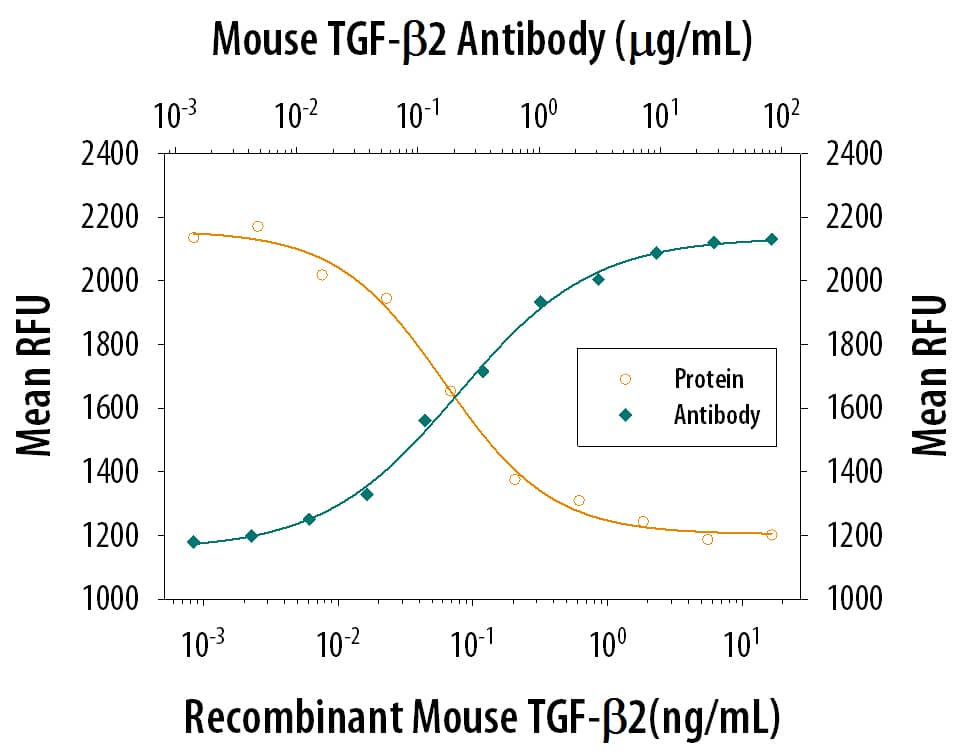
TGF‑ beta2 Inhibition of IL‑4-dependent Cell Proliferation and Neutralization by Mouse TGF‑ beta2 Antibody.
Recombinant Mouse TGF-beta 2 (Catalog # 7346-B2) inhibits Recombinant Mouse IL-4 (Catalog # 404-ML) induced proliferation in the HT-2 mouse T cell line in a dose-dependent manner (orange line). Inhibition of Recombinant Mouse IL-4 (7.5 ng/mL) activity elicited by Recombinant Mouse TGF-beta 2 (5 ng/mL) is neutralized (green line) by increasing concen-trations of Rat Anti-Mouse TGF-beta 2 Monoclonal Antibody (Catalog # MAB7346). The ND50 is typically 0.15-0.75 ug/mL.Applications for Mouse TGF-beta 2 Antibody
Application
Recommended Usage
Neutralization
Measured by its ability to neutralize TGF‑ beta2 inhibition of IL-4-dependent proliferation in the HT‑2 mouse T cell line. Tsang, M. et al. (1995) Cytokine 7:389. The Neutralization Dose (ND50) is typically 0.15-0.75 ug/mL in the presence of 5 ng/mL Recombinant Mouse TGF‑ beta2 and 7.5 ng/mL Recombinant Mouse IL-4.
Formulation, Preparation, and Storage
Purification
Protein A or G purified from hybridoma culture supernatant
Reconstitution
Sterile PBS to a final concentration of 0.5 mg/mL. For liquid material, refer to CoA for concentration.
Formulation
Lyophilized from a 0.2 μm filtered solution in PBS with Trehalose. *Small pack size (SP) is supplied either lyophilized or as a 0.2 µm filtered solution in PBS.
Shipping
Lyophilized product is shipped at ambient temperature. Liquid small pack size (-SP) is shipped with polar packs. Upon receipt, store immediately at the temperature recommended below.
Stability & Storage
Use a manual defrost freezer and avoid repeated freeze-thaw cycles.
- 12 months from date of receipt, -20 to -70 °C as supplied.
- 1 month, 2 to 8 °C under sterile conditions after reconstitution.
- 6 months, -20 to -70 °C under sterile conditions after reconstitution.
Background: TGF-beta 2
Long Name
Transforming Growth Factor beta 2
Alternate Names
TGFB2, TGFbeta 2
Gene Symbol
TGFB2
UniProt
Additional TGF-beta 2 Products
Product Documents for Mouse TGF-beta 2 Antibody
Product Specific Notices for Mouse TGF-beta 2 Antibody
For research use only
Loading...
Loading...
Loading...