Mouse TLR2 Antibody
R&D Systems, part of Bio-Techne | Catalog # MAB1530


Key Product Details
Validated by
Species Reactivity
Validated:
Cited:
Applications
Validated:
Cited:
Label
Antibody Source
Product Specifications
Immunogen
Gln25-Leu590
Accession # Q9QUN7
Specificity
Clonality
Host
Isotype
Scientific Data Images for Mouse TLR2 Antibody
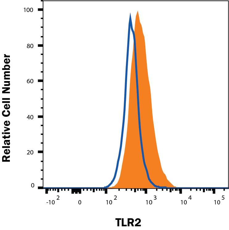
Detection of TLR2 in Raw264.7 cells by Flow Cytometry.
Raw264.7 cells stimulated with 100 ng/mL LPS overnight were stained with Rat Anti-Mouse TLR2 Monoclonal Antibody (Catalog # MAB1530, filled histogram) or isotype control antibody (Catalog # MAB0061, open histogram), followed by Fluorescein-conjugated Anti-Rat IgG Secondary Antibody (Catalog # F0104B). View our protocol for Staining Membrane-associated Proteins.Detection of Mouse TLR2 by Western Blot
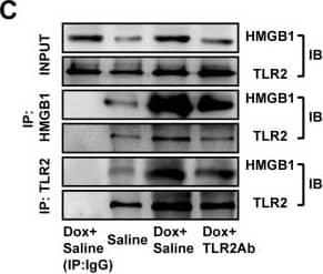
TLR2Ab suppressed the expression of endogenous TLR2 agonists and interaction of TLR2 with HMGB1.(A) Expression of HMGB1 and Hsp70 were reduced in TLR2Ab-treated mice, but not in TLR4Ab group. (B) TLR2Ab and TLR4Ab suppressed its downstream signaling pathway. Data are shown as mean ± SEM (n = 5/group). (C) TLR2Ab inhibited the interaction of TLR2 with HMGB1 as indicated by co-immunoprecipitation and western blot. Data are representatives of two experiments with identical results. (D) TLR2Ab decreased the co-localization of TLR2 and HMGB1 as shown by confocal microscopy. Data are representatives of two experiments with identical results. #p<0.05, ##p<0.01, ###p<0.001 versus saline-treated group; *p<0.05, **p<0.01, ***p<0.001 versus Dox+IgG-treated group. Image collected and cropped by CiteAb from the following publication (https://pubmed.ncbi.nlm.nih.gov/22808256), licensed under a CC-BY license. Not internally tested by R&D Systems.Detection of Mouse TLR2 by Western Blot
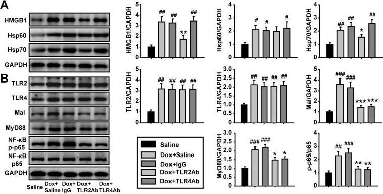
TLR2Ab suppressed the expression of endogenous TLR2 agonists and interaction of TLR2 with HMGB1.(A) Expression of HMGB1 and Hsp70 were reduced in TLR2Ab-treated mice, but not in TLR4Ab group. (B) TLR2Ab and TLR4Ab suppressed its downstream signaling pathway. Data are shown as mean ± SEM (n = 5/group). (C) TLR2Ab inhibited the interaction of TLR2 with HMGB1 as indicated by co-immunoprecipitation and western blot. Data are representatives of two experiments with identical results. (D) TLR2Ab decreased the co-localization of TLR2 and HMGB1 as shown by confocal microscopy. Data are representatives of two experiments with identical results. #p<0.05, ##p<0.01, ###p<0.001 versus saline-treated group; *p<0.05, **p<0.01, ***p<0.001 versus Dox+IgG-treated group. Image collected and cropped by CiteAb from the following publication (https://pubmed.ncbi.nlm.nih.gov/22808256), licensed under a CC-BY license. Not internally tested by R&D Systems.Applications for Mouse TLR2 Antibody
CyTOF-ready
Flow Cytometry
Sample: Raw264.7 cells stimulated with 100 ng/mL LPS overnight
Reviewed Applications
Read 2 reviews rated 4.5 using MAB1530 in the following applications:
Formulation, Preparation, and Storage
Purification
Reconstitution
Formulation
Shipping
Stability & Storage
- 12 months from date of receipt, -20 to -70 °C as supplied.
- 1 month, 2 to 8 °C under sterile conditions after reconstitution.
- 6 months, -20 to -70 °C under sterile conditions after reconstitution.
Background: TLR2
The Toll-like family of molecules are a group of integral membrane proteins that serve as pattern recognition receptors for microbial pathogens (1-4). To date, there are at least eleven mouse and ten human members that activate the innate immune system following exposure to a variety of microbial species (1, 3). All Toll-like receptors (TLRs) are type I transmembrane (TM) proteins that exist either in the plasma membrane or in the membranes of endosomal structures (where they bind intracellular nucleic acids) (3). All TLRs also contain a large number of extracellular leucine-rich repeats (LRRs) and a cytoplasmic tail with a Toll/IL-1 receptor (TIR) domain. Mouse Toll-like receptor-2 (TLR2) is a 97 kDa, 760 amino acid (aa) glycoprotein that contains a 563 aa extracellular region, a 21 aa TM segment, and a 176 aa cytoplasmic domain (5, 6). The extracellular region contains 16 leucine-rich repeats, while the cytoplasmic tail shows one 146 aa TIR domain. The receptor is expressed on a number of cell types including T cells ( alpha beta and gamma delta), monocytes, dendritic cells, neutrophils, B cells, endothelial cells, mast cells, NK cells, macrophages, and hepatocytes (1, 4, 5, 7, 8). TLR2 functions as part of a heterodimeric complex with either TLR1 or TLR6 (1, 3, 4). These complexes recognize lipoproteins and glycolipids from gram-positive and gram-negative bacteria as well as mycoplasma and yeast. TLR2/TLR1 heterodimers recognize triacylated lipopeptides from a variety of microorganisms. The TLR2/TLR6 heterodimer preferentially recognizes diacylated lipopeptides (9). Biglycan is also known to activate TLR2, but the context is unclear (8). Notably, in human, TLR2 also dimerizes with TLR10. But the TLR10 gene in mouse (but not rat) is mutationally inactive, and thus this complex is nonfunctional (10). Upon ligand recognition, TLR2 delivers an activating signal via the associated adapter molecules, MyD88 and TIRAP (1, 11). Activation via TLR2 also results in production of a number of pro-inflammatory cytokines including TNF-alpha, IL-2, IL-6, IL-12, and MIP-2 (1, 3). The extracellular region of mouse TLR2 is 89%, 67%, 81%, and 65% aa identical to the equivalent region in rat, human, hamster, and canine, respectively.
References
- Wetzler, L. (2003) Vaccine 21:S2/55.
- Netea, M. et al. (2004) J. Leukoc. Biol. 75:749.
- Dunne, A. and L. O’Neill (2005) FEBS. Lett. 579:3330.
- Hopkins, P.A. and S. Sriskandan (2005) Clin. Exp. Immunol. 140:395.
- Matsuguchi, T. et al. (2000) Blood 95:1378.
- Meng, G. et al. (2005) Immunol. Lett. 98:200.
- Flo, T. et al. (2001) J. Leukoc. Biol. 69:474.
- Schaefer, L. et al. (2005) J. Clin. Invest. 115:2223.
- Akira, S. (2003) Curr. Opin. Immunol. 15:5.
- Hasan, U. et al. (2005) J. Immunol. 174:2942.
- Yamamoto M. et al. (2002) Nature 420:324.
Long Name
Alternate Names
Gene Symbol
UniProt
Additional TLR2 Products
Product Documents for Mouse TLR2 Antibody
Product Specific Notices for Mouse TLR2 Antibody
For research use only

