Mouse TNF-alpha Antibody
R&D Systems, part of Bio-Techne | Catalog # AB-410-NA

Key Product Details
Species Reactivity
Validated:
Mouse
Cited:
Mouse, Transgenic Mouse
Applications
Validated:
Neutralization, Western Blot
Cited:
Cell Culture, Immunohistochemistry, Neutralization, Western Blot
Label
Unconjugated
Antibody Source
Polyclonal Goat IgG
Product Specifications
Immunogen
E. coli-derived recombinant mouse TNF-alpha
Leu80-Leu235
Accession # P06804
Leu80-Leu235
Accession # P06804
Specificity
Detects mouse TNF-alpha in direct ELISAs and Western blots. In direct ELISAs, approximately 15% cross-reactivity with human TNF‑ alpha is observed, and less than 5% cross-reactivity with recombinant bovine TNF‑ alpha, recombinant canine TNF‑ alpha, recombinant equine TNF‑ alpha, recombinant feline TNF‑ alpha, and recombinant porcine TNF‑ alpha is observed.
Clonality
Polyclonal
Host
Goat
Isotype
IgG
Endotoxin Level
<0.10 EU per 1 μg of the antibody by the LAL method.
Scientific Data Images for Mouse TNF-alpha Antibody
Cytotoxicity Induced by TNF-alpha and Neutralization by Mouse TNF-alpha Antibody.
Recombinant Mouse TNF-a (Catalog # 410-MT) induces cytotoxicity in the the L-929 mouse fibroblast cell line in a dose-dependent manner (orange line), as measured by crystal violet staining. Cytotoxicity elicited by Recombinant Mouse TNF-a (0.25 ng/mL) is neutralized (green line) by increasing concentrations of Mouse TNF-a Polyclonal Antibody (Catalog # AB-410-NA). The ND50 is typically 0.2-0.6 µg/mL in the presence of the metabolic inhibitor actinomycin D (1 µg/mL).Detection of Mouse Mouse TNF-alpha Antibody by Western Blot
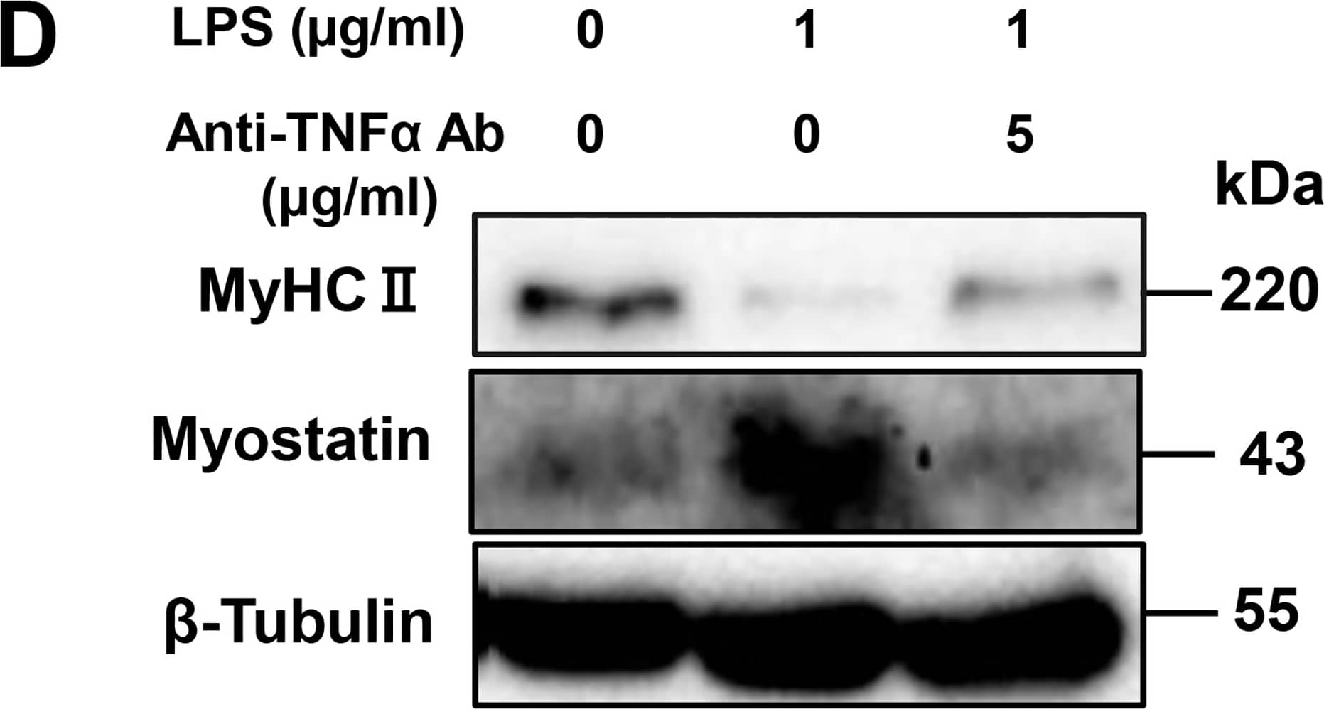
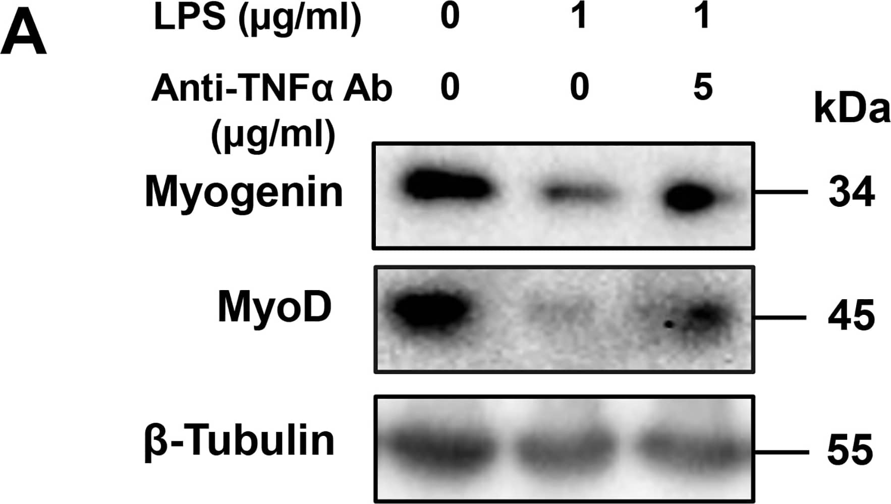
Effect of a TNF-alpha -neutralizing antibody on LPS-induced perturbation of muscle differentiation.(A) C2C12 myoblasts were cultured for 48 h in DM alone, LPS (1 μg/mL), or LPS (1 μg/mL) plus TNF-alpha -neutralizing antibody (5 μg/mL). A representative western blot probed with antibodies to myogenin, MyoD, or beta-tubulin (internal standard) is shown. (B and C) Quantification of the data presented in (A). Data are the mean ± SEM of 7–15 independent experiments. (D) C2C12 myoblasts were cultured for 144 h as described in (A). A representative western blot probed with antibodies to MyHC II, myostatin, or beta-tubulin (internal standard) is shown. (E and F) Quantification of the data presented in (D). Data are the mean ± SEM of 5–21 independent experiments. (G) Cells were treated as described in (A), and NF-kappa B activity was analyzed by ELISA. Data are the mean ± SEM of 7 independent experiments performed in duplicate. ***p < 0.001, **p < 0.01, *p < 0.05, ###p < 0.001, ##p < 0.01, #p < 0.05 by one-way ANOVA followed by Tukey’s honest significant difference test. Image collected and cropped by CiteAb from the following publication (https://pubmed.ncbi.nlm.nih.gov/28742154), licensed under a CC-BY license. Not internally tested by R&D Systems.Detection of Mouse Mouse TNF-alpha Antibody by Western Blot
Effect of a TNF-alpha -neutralizing antibody on LPS-induced perturbation of muscle differentiation.(A) C2C12 myoblasts were cultured for 48 h in DM alone, LPS (1 μg/mL), or LPS (1 μg/mL) plus TNF-alpha -neutralizing antibody (5 μg/mL). A representative western blot probed with antibodies to myogenin, MyoD, or beta-tubulin (internal standard) is shown. (B and C) Quantification of the data presented in (A). Data are the mean ± SEM of 7–15 independent experiments. (D) C2C12 myoblasts were cultured for 144 h as described in (A). A representative western blot probed with antibodies to MyHC II, myostatin, or beta-tubulin (internal standard) is shown. (E and F) Quantification of the data presented in (D). Data are the mean ± SEM of 5–21 independent experiments. (G) Cells were treated as described in (A), and NF-kappa B activity was analyzed by ELISA. Data are the mean ± SEM of 7 independent experiments performed in duplicate. ***p < 0.001, **p < 0.01, *p < 0.05, ###p < 0.001, ##p < 0.01, #p < 0.05 by one-way ANOVA followed by Tukey’s honest significant difference test. Image collected and cropped by CiteAb from the following publication (https://pubmed.ncbi.nlm.nih.gov/28742154), licensed under a CC-BY license. Not internally tested by R&D Systems.Applications for Mouse TNF-alpha Antibody
Application
Recommended Usage
Western Blot
1 µg/mL
Sample: Recombinant Mouse TNF-alpha (Catalog # 410-MT)
Sample: Recombinant Mouse TNF-alpha (Catalog # 410-MT)
Neutralization
Measured by its ability to neutralize TNF‑ alpha-induced cytotoxicity in the L‑929 mouse fibroblast cell line [Matthews, N. and M.L. Neale (1987) in Lymphokines and Interferons, A Practical Approach. Clemens, M.J. et al. (eds): IRL Press. 221]. The Neutralization Dose (ND50) is typically 0.2-0.6 µg/mL in the presence of 0.25 ng/mL Recombinant Mouse TNF‑ alpha and 1 µg/mL actinomycin D.
Formulation, Preparation, and Storage
Purification
Protein A or G purified
Reconstitution
Reconstitute at 1 mg/mL in sterile PBS.
Formulation
Lyophilized from a 0.2 μm filtered solution in PBS with Trehalose.
Shipping
The product is shipped at ambient temperature. Upon receipt, store it immediately at the temperature recommended below.
Stability & Storage
Use a manual defrost freezer and avoid repeated freeze-thaw cycles.
- 12 months from date of receipt, -20 to -70 °C as supplied.
- 1 month, 2 to 8 °C under sterile conditions after reconstitution.
- 6 months, -20 to -70 °C under sterile conditions after reconstitution.
Background: TNF-alpha
Long Name
Tumor Necrosis Factor alpha
Alternate Names
Cachetin, DIF, TNF, TNF-A, TNFA, TNFalpha, TNFG1F, TNFSF1A, TNFSF2
Entrez Gene IDs
Gene Symbol
TNF
UniProt
Additional TNF-alpha Products
Product Documents for Mouse TNF-alpha Antibody
Product Specific Notices for Mouse TNF-alpha Antibody
For research use only
Loading...
Loading...
Loading...
Loading...
Loading...


