Mouse TNF RII/TNFRSF1B APC-conjugated Antibody
R&D Systems, part of Bio-Techne | Catalog # FAB426A


Key Product Details
Species Reactivity
Validated:
Cited:
Applications
Validated:
Cited:
Label
Antibody Source
Product Specifications
Immunogen
Extracellular domain
Specificity
Clonality
Host
Isotype
Scientific Data Images for Mouse TNF RII/TNFRSF1B APC-conjugated Antibody
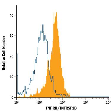
Detection of TNF RII/TNFRSF1B in L-929 Mouse Cell Line by Flow Cytometry.
L‑929 mouse fibroblast cell line was stained with Hamster Anti-Mouse TNF RII/TNFRSF1B APC‑conjugated Monoclonal Antibody (Catalog # FAB426A, filled histogram) or Hamster IgG Allophycocyanin Control Antibody (open histogram). View our protocol for Staining Membrane-associated Proteins.Applications for Mouse TNF RII/TNFRSF1B APC-conjugated Antibody
Flow Cytometry
Sample: L‑929 mouse fibroblast cell line
Formulation, Preparation, and Storage
Purification
Formulation
Shipping
Stability & Storage
- 12 months from date of receipt, 2 to 8 °C as supplied.
Background: TNF RII/TNFRSF1B
Two types of soluble TNF receptors have been identified in human serum and urine which can neutralize the biological activities of TNF-alpha and TNF-beta. These binding proteins represent truncated forms of the two types of high-affinity cell surface receptors for TNF (TNFR-p60 Type B and TNFR-p80 Type A). Soluble TNF RII corresponds to TNFR-p80 Type A. In the new TNF superfamily nomenclature, TNF RII is referred to as TNFRSF1B. These apparent soluble forms of the receptors appear to arise as a result of shedding of the extracellular domains of the membrane-bound receptors. Normal concentrations as high as 4 ng/mL are found in the serum of healthy individuals, and even higher levels may be found in some pathological conditions. Although the physiological role of these proteins is not known, it has been speculated that shedding of the soluble receptors in response to TNF release could serve as a mechanism to scavenge the TNF not immediately bound and thus localize the inflammatory response. It is also possible that the pool of TNF bound to soluble receptors could represent a reservoir for the controlled release of TNF.
Long Name
Alternate Names
Gene Symbol
Additional TNF RII/TNFRSF1B Products
Product Documents for Mouse TNF RII/TNFRSF1B APC-conjugated Antibody
Product Specific Notices for Mouse TNF RII/TNFRSF1B APC-conjugated Antibody
For research use only