Mouse VCAM-1/CD106 Antibody
R&D Systems, part of Bio-Techne | Catalog # MAB6434

Key Product Details
Validated by
Species Reactivity
Validated:
Cited:
Applications
Validated:
Cited:
Label
Antibody Source
Product Specifications
Immunogen
Phe25-Glu698
Accession # P29533
Specificity
Clonality
Host
Isotype
Scientific Data Images for Mouse VCAM-1/CD106 Antibody
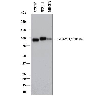
Detection of Mouse VCAM‑1/CD106 by Western Blot.
Western blot shows lysates of 3T3-L1 mouse embryonic fibroblast adipose-like cell line, C2C12 mouse myoblast cell line, and NIH-3T3 mouse embryonic fibroblast cell line. PVDF membrane was probed with 2 µg/mL of Rat Anti-Mouse VCAM-1/CD106 Monoclonal Antibody (Catalog # MAB6434) followed by HRP-conjugated Anti-Rat IgG Secondary Antibody (Catalog # HAF005). A specific band was detected for VCAM-1/ CD106 at approximately 95 kDa (as indicated). This experiment was conducted under reducing conditions and using Immunoblot Buffer Group 1.Detection of Mouse VCAM‑1/CD106 by Simple WesternTM.
Simple Western lane view shows lysate of 3T3-L1 mouse embryonic fibroblast adipose-like cell line, loaded at 0.2 mg/mL. A specific band was detected for VCAM-1/CD106 at approximately 117 kDa (as indicated) using 100 µg/mL of Rat Anti-Mouse VCAM-1/CD106 Monoclonal Antibody (Catalog # MAB6434) followed by 1:50 dilution of HRP-conjugated Anti-Rat IgG Secondary Antibody (Catalog # HAF005). This experiment was conducted under reducing conditions and using the 12-230 kDa separation system.Detection of Mouse VCAM-1/CD106 by Western Blot
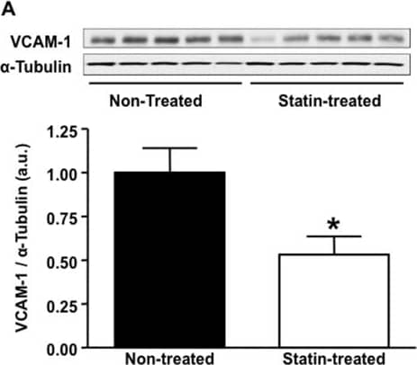
Effect of statin treatment on aortic expression of VCAM-1.(A) VCAM-1 protein expression in the ascending aorta assessed by Western blot in non-treated (lanes 1–5) versus statin treated (lanes 6–10) animals, n = 5 per group, *p<0.01 vs non-treated animals. Representative examples of fluorescent immunohistochemistry images of the base of the aorta demonstrating endothelial VCAM-1 expression (red fluorescence) in a non-treated animal (B) and a statin treated animal (C), the green fluorescence is autofluorescence delineating vessel anatomy. Image collected and cropped by CiteAb from the following publication (https://pubmed.ncbi.nlm.nih.gov/23554922), licensed under a CC-BY license. Not internally tested by R&D Systems.Applications for Mouse VCAM-1/CD106 Antibody
Simple Western
Sample: C2C12 mouse myoblast cell line, 3T3‑L1 mouse embryonic fibroblast adipose-like cell line, and NIH‑3T3 mouse embryonic fibroblast cell line
Western Blot
Sample: 3T3‑L1 mouse embryonic fibroblast adipose-like cell line, C2C12 mouse myoblast cell line, and NIH‑3T3 mouse embryonic fibroblast cell line
Formulation, Preparation, and Storage
Purification
Reconstitution
Formulation
Shipping
Stability & Storage
- 12 months from date of receipt, -20 to -70 °C as supplied.
- 1 month, 2 to 8 °C under sterile conditions after reconstitution.
- 6 months, -20 to -70 °C under sterile conditions after reconstitution.
Background: VCAM-1/CD106
VCAM-1 (CD106), a member of the immunoglobulin superfamily, is a cell surface protein expressed by activated endothelial cells and certain leukocytes (such as macrophages). VCAM-1 expression is induced by IL-1 beta, IL-4, TNF-alpha, and IFN-gamma. VCAM-1 binds to leukocyte integrins VLA-4 and alpha4 beta7. The human and mouse VCAM-1 proteins share approximately 76% amino acid similarity.
During the inflammatory adhesion mechanism, activated integrins halt rolling leukocytes and attach them firmly to the vascular endothelium. They do this by binding to their ligands, for example VCAM-1, on endothelium. The VCAM-1: VLA-4/ alpha4 beta7 interaction is also thought to be involved in the extravasation of white blood cells through the blood vessel wall to sites of inflammation.
ELISA techniques have shown that detectable levels of soluble VCAM-1 are present in the biological fluids of apparently normal individuals. Furthermore, a number of studies have reported that levels of VCAM-1 may be elevated or lowered in subjects with a variety of pathological conditions.
Long Name
Alternate Names
Gene Symbol
UniProt
Additional VCAM-1/CD106 Products
Product Documents for Mouse VCAM-1/CD106 Antibody
Product Specific Notices for Mouse VCAM-1/CD106 Antibody
For research use only


