Detection of Mouse Mouse VEGFR1/Flt-1 Antibody by Immunohistochemistry
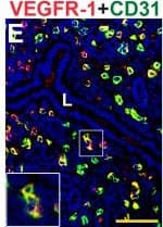
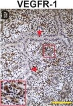
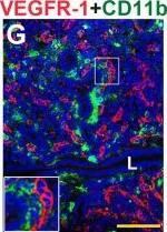
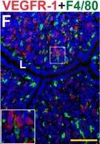
VEGFR-1, CD31, F4/80, and CD11b expression in the pre-implantation mouse uterus. IHC and double staining IF were performed on E3.5 uterine cross-sections. (A) Schematic representation of an E3.5 mouse uterus showing lumen (arrowheads), glands, stroma (s), and myometrium (myo). (B) ECs, detected by CD31 staining (brown), are observed throughout the stroma and myometrium, similar to the non-pregnant state. (C) Macrophages, detected by F4/80 staining (brown), are observed throughout the stroma and are abundant adjacent to the lumen and glands at E3.5. (D) VEGFR-1+ cells (brown), are distributed throughout the stroma and cell associated VEGFR-1 expression highlighted in the inset. (E) Double staining for VEGFR-1 (red) and CD31 (green) demonstrates expression of VEGFR-1 on CD31+ ECs throughout the stroma. (F) VEGFR-1+ cells (red) and F4/80+ macrophages (green) are distributed throughout the stroma. VEGFR-1 and F4/80 co-expression is not observed. (G) VEGFR-1+ cells (red) and CD11b+ monocytes (green) are distributed throughout the stroma. VEGFR-1 and CD11b co-expression is not observed. L, lumen. Scale bars B, C = 100 μm. Scale bars D – F = 50 μm. Image collected and cropped by CiteAb from the following publication (https://pubmed.ncbi.nlm.nih.gov/25101167), licensed under a CC-BY license. Not internally tested by R&D Systems.
Detection of Mouse Mouse VEGFR1/Flt-1 Antibody by Immunohistochemistry
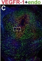
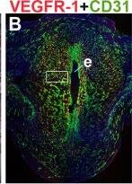
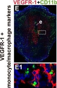
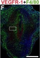
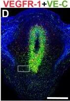
VEGFR-1 expression in endothelial cells and macrophages in the post-implantation uterus. H&E and double staining IF were performed on E6.5 frontal uterine sections. (A) H&E of a post-implantation mouse uterus showing the embryo (e), anti-mesometrial (am) and mesometrial (m) areas. (B-F) VEGFR-1+ cells (red) are observed in the decidua, with abundant expression in the primary decidual zone surrounding the implanted embryo. (B) Double-staining for VEGFR-1+ (red) and CD31+ (green) cells demonstrates expression of VEGFR-1 in a subset of CD31+ ECs. (C) Double-staining for VEGFR-1+ (red) and endomucin+ (green) cells demonstrates expression of VEGFR-1 in a subset of endomucin+ ECs. (D) Double-staining for VEGFR-1+ (red) and VE-cadherin+ (green) cells demonstrates expression of VEGFR-1 in VE-cadherin+ ECs. (E) Double-staining for VEGFR-1+ (red) and CD11b+ (green) cells demonstrates that VEGFR-1 is not expressed in CD11b+ monocytes. (F) Double-staining for VEGFR-1+ (red) and F4/80+ (green) cells demonstrates that VEGFR-1 is not expressed in F4/80+ macrophages. VEGFR-1+ cells are adjacent to CD11b+ monocytes and F4/80+ macrophages. White boxes in (B-F) indicate areas of the uteri magnified below (B1-F1). (A-F) Scale bar = 500 μm. (B1-F1) Scale bar = 20 μm. Image collected and cropped by CiteAb from the following publication (https://pubmed.ncbi.nlm.nih.gov/25101167), licensed under a CC-BY license. Not internally tested by R&D Systems.
Detection of Mouse Mouse VEGFR1/Flt-1 Antibody by Immunohistochemistry
VEGFR-1, CD31, F4/80, and CD11b expression in the pre-implantation mouse uterus. IHC and double staining IF were performed on E3.5 uterine cross-sections. (A) Schematic representation of an E3.5 mouse uterus showing lumen (arrowheads), glands, stroma (s), and myometrium (myo). (B) ECs, detected by CD31 staining (brown), are observed throughout the stroma and myometrium, similar to the non-pregnant state. (C) Macrophages, detected by F4/80 staining (brown), are observed throughout the stroma and are abundant adjacent to the lumen and glands at E3.5. (D) VEGFR-1+ cells (brown), are distributed throughout the stroma and cell associated VEGFR-1 expression highlighted in the inset. (E) Double staining for VEGFR-1 (red) and CD31 (green) demonstrates expression of VEGFR-1 on CD31+ ECs throughout the stroma. (F) VEGFR-1+ cells (red) and F4/80+ macrophages (green) are distributed throughout the stroma. VEGFR-1 and F4/80 co-expression is not observed. (G) VEGFR-1+ cells (red) and CD11b+ monocytes (green) are distributed throughout the stroma. VEGFR-1 and CD11b co-expression is not observed. L, lumen. Scale bars B, C = 100 μm. Scale bars D – F = 50 μm. Image collected and cropped by CiteAb from the following publication (https://pubmed.ncbi.nlm.nih.gov/25101167), licensed under a CC-BY license. Not internally tested by R&D Systems.
Detection of Mouse Mouse VEGFR1/Flt-1 Antibody by Immunohistochemistry
VEGFR-1 expression in endothelial cells and macrophages in the post-implantation uterus. H&E and double staining IF were performed on E6.5 frontal uterine sections. (A) H&E of a post-implantation mouse uterus showing the embryo (e), anti-mesometrial (am) and mesometrial (m) areas. (B-F) VEGFR-1+ cells (red) are observed in the decidua, with abundant expression in the primary decidual zone surrounding the implanted embryo. (B) Double-staining for VEGFR-1+ (red) and CD31+ (green) cells demonstrates expression of VEGFR-1 in a subset of CD31+ ECs. (C) Double-staining for VEGFR-1+ (red) and endomucin+ (green) cells demonstrates expression of VEGFR-1 in a subset of endomucin+ ECs. (D) Double-staining for VEGFR-1+ (red) and VE-cadherin+ (green) cells demonstrates expression of VEGFR-1 in VE-cadherin+ ECs. (E) Double-staining for VEGFR-1+ (red) and CD11b+ (green) cells demonstrates that VEGFR-1 is not expressed in CD11b+ monocytes. (F) Double-staining for VEGFR-1+ (red) and F4/80+ (green) cells demonstrates that VEGFR-1 is not expressed in F4/80+ macrophages. VEGFR-1+ cells are adjacent to CD11b+ monocytes and F4/80+ macrophages. White boxes in (B-F) indicate areas of the uteri magnified below (B1-F1). (A-F) Scale bar = 500 μm. (B1-F1) Scale bar = 20 μm. Image collected and cropped by CiteAb from the following publication (https://pubmed.ncbi.nlm.nih.gov/25101167), licensed under a CC-BY license. Not internally tested by R&D Systems.


