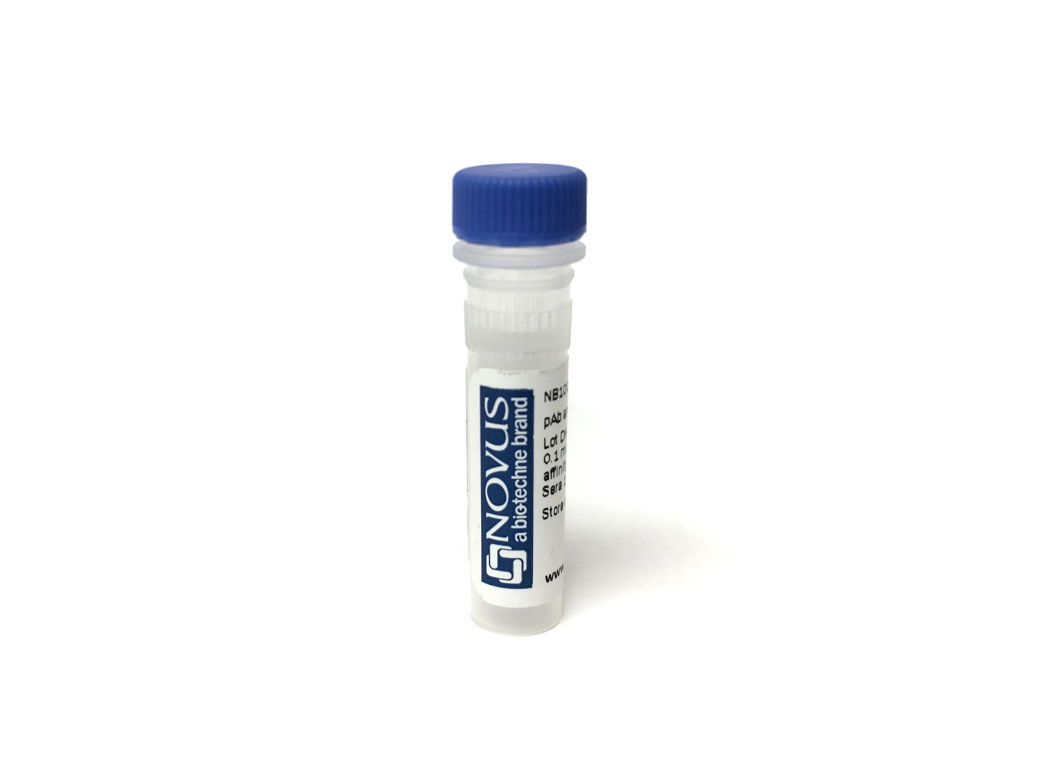
P-Cadherin Antibody (MM0508-9V11) [PE/Cy7]
Novus Biologicals, part of Bio-Techne | Catalog # NBP2-11842PECY7


Conjugate
Catalog #
Forumulation
Catalog #
Key Product Details
Species Reactivity
Human
Applications
Flow Cytometry
Label
PE/Cy7 (Excitation = 488 nm, Emission = 778 nm)
Antibody Source
Monoclonal Mouse IgG1 Clone # MM0508-9V11
Concentration
Please see the vial label for concentration. If unlisted please contact technical services.
Product Specifications
Immunogen
Human recombinant P-Cadherin (extracellular domain)
Marker
Intercellular Junctions Marker
Clonality
Monoclonal
Host
Mouse
Isotype
IgG1
Applications for P-Cadherin Antibody (MM0508-9V11) [PE/Cy7]
Application
Recommended Usage
Flow Cytometry
Optimal dilutions of this antibody should be experimentally determined.
Application Notes
Optimal dilution of this antibody should be experimentally determined. For optimal results using our Tandem dyes, please avoid prolonged exposure to light or extreme temperature fluctuations. These can lead to irreversible degradation or decoupling. When staining intracellular targets, specific attention to the fixation and permeabilization steps in your flow protocol may be required. Please contact our technical support team at technical@novusbio.com if you have any questions.
Formulation, Preparation, and Storage
Purification
Protein G purified
Formulation
PBS
Preservative
0.05% Sodium Azide
Concentration
Please see the vial label for concentration. If unlisted please contact technical services.
Shipping
The product is shipped with polar packs. Upon receipt, store it immediately at the temperature recommended below.
Stability & Storage
Store at 4C in the dark. Do not freeze.
Background: P-Cadherin
Long Name
Placental Cadherin
Alternate Names
CAD3, Cadherin-3, CDH3, CDHP, HJMD, PCAD, PCadherin
Gene Symbol
CDH3
Additional P-Cadherin Products
Product Documents for P-Cadherin Antibody (MM0508-9V11) [PE/Cy7]
Product Specific Notices for P-Cadherin Antibody (MM0508-9V11) [PE/Cy7]
This product is for research use only and is not approved for use in humans or in clinical diagnosis. Primary Antibodies are guaranteed for 1 year from date of receipt.
Loading...
Loading...
Loading...
Loading...
Loading...
Loading...