p53 Antibody - BSA Free
Novus Biologicals, part of Bio-Techne | Catalog # NBP2-19667

Key Product Details
Validated by
Species Reactivity
Applications
Label
Antibody Source
Format
Concentration
Product Specifications
Immunogen
Localization
Clonality
Host
Isotype
Theoretical MW
Disclaimer note: The observed molecular weight of the protein may vary from the listed predicted molecular weight due to post translational modifications, post translation cleavages, relative charges, and other experimental factors.
Scientific Data Images for p53 Antibody - BSA Free
p53 antibody [N1], N-term detects p53 protein at nucleus by immunofluorescent analysis.
Samples: HCT 116 cells mock (left) and treated with 30 uM Cisplatin for 24 hrs (right) were fixed in 4% paraformaldehyde at RT for 15 min.
Green: p53 protein stained by p53 antibody [N1], N-term.
Blue: Hoechst 33342 staining.
Scale bar = 10 um.
Western Blot: p53 Antibody [NBP2-19667] - p53 antibody [N1], N-term detects p53 protein at nucleus by immunofluorescent analysis.Samples: HCT 116 cells mock (left) and treated with 30 uM Cisplatin for 24 hrs (right) were fixed in 4% paraformaldehyde at RT for 15 min.
Green: p53 protein stained by p53 antibody [N1], N-term.
Blue: Hoechst 33342 staining.
Scale bar = 10 um.
Immunocytochemistry/ Immunofluorescence: p53 Antibody [NBP2-19667]
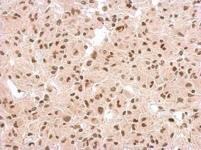
Immunocytochemistry/Immunofluorescence: p53 Antibody [NBP2-19667] - Confocal immunofluorescence analysis of paraformaldehyde-fixed U2OS, using p53 antibody (Green) at 1:500 dilution. Alpha-tubulin filaments are labeled with Alpha-tubulin antibody (Red) at 1:2000.U373 xenograft, using p53 antibody at 1:500 dilution. Antigen Retrieval: Trilogy™ (EDTA based, pH 8.0) buffer, 15min
U373 xenograft, using p53 antibody at 1:500 dilution. Antigen Retrieval: Trilogy™ (EDTA based, pH 8.0) buffer, 15minApplications for p53 Antibody - BSA Free
Immunocytochemistry/ Immunofluorescence
Immunohistochemistry
Immunohistochemistry-Paraffin
Immunoprecipitation
Western Blot
Reviewed Applications
Read 1 review rated 5 using NBP2-19667 in the following applications:
Formulation, Preparation, and Storage
Purification
Formulation
Format
Preservative
Concentration
Shipping
Stability & Storage
Background: p53
Additional p53 Products
Product Documents for p53 Antibody - BSA Free
Product Specific Notices for p53 Antibody - BSA Free
This product is for research use only and is not approved for use in humans or in clinical diagnosis. Primary Antibodies are guaranteed for 1 year from date of receipt.
⚠ WARNING: This product can expose you to chemicals including mercury, which is known to the State of California to cause reproductive toxicity with developmental effects. For more information go to www.P65Warnings.ca.gov.
![Immunocytochemistry/ Immunofluorescence: p53 Antibody [NBP2-19667] Immunocytochemistry/ Immunofluorescence: p53 Antibody [NBP2-19667]](https://resources.bio-techne.com/images/products/p53-Antibody-Immunocytochemistry-Immunofluorescence-NBP2-19667-img0003.jpg)

![Immunoprecipitation: p53 Antibody [NBP2-19667] Immunoprecipitation: p53 Antibody [NBP2-19667]](https://resources.bio-techne.com/images/products/p53-Antibody-Immunoprecipitation-NBP2-19667-img0007.jpg)
![Whole cell extract (30 μg) were separated by 10% SDS-PAGE, and the membrane was blotted with p53 antibody [N1], N-term diluted at 1:1000. The HRP-conjugated anti-rabbit IgG antibody (NBP2-19301) was used to detect the primary antibody.](https://resources.bio-techne.com/images/products/antibody/nbp2-19667_rabbit-polyclonal-p53-antibody-western-blot-22320239226.jpg)
![p53 antibody [N1], N-term detects p53 protein at nucleus by immunofluorescent analysis.<br></br>Samples: HCT 116 cells mock (left) and treated with 30 uM Cisplatin for 24 hrs (right) were fixed in 4% paraformaldehyde at RT for 15 min.<br>Green: p53 protein stained by p53 antibody [N1], N-term.<br>Blue: Hoechst 33342 staining.<br>Scale bar = 10 um.](https://resources.bio-techne.com/images/products/p53-Antibody-Western-Blot-NBP2-19667-img0006.jpg)