Phospho-GAP‑43 (S41) Antibody
R&D Systems, part of Bio-Techne | Catalog # PPS006

Key Product Details
Species Reactivity
Validated:
Human, Mouse, Rat, Bovine, Canine, Chicken, Primate, Xenopus, Zebrafish
Cited:
Mouse
Applications
Validated:
Western Blot
Cited:
Western Blot
Label
Unconjugated
Antibody Source
Polyclonal Rabbit IgG
Product Specifications
Immunogen
Phosphopeptide corresponding to amino acid residues surrounding the phospho-S41 of GAP-43
Specificity
Specific for the ~50 kDa GAP-43 protein phosphorylated at S41 in Western blots. In some tissues the antibody also recognizes a higher molecular weight protein, that is also recognized by the Pan GAP-43 antibody. This may be a GAP-43 aggregate or oligomer.
Clonality
Polyclonal
Host
Rabbit
Isotype
IgG
Scientific Data Images for Phospho-GAP‑43 (S41) Antibody
Detection of Phospho-GAP‑43 (S41) by Western blot.
Western blot of rat cortex lysate showing specific immunolabeling of the ~50 kDa GAP-43 protein phosphorylated at S41 (Control). The phosphospecificity of this labeling is demonstrated by treatment with 1200 U of lambda Phosphatase ( lambda-PPase) for 30 minutes before being exposed to the Anti-Phospho-GAP-43 (S41). The immunolabeling is completely eliminated by treatment with lambda-PPase.Detection of Mouse GAP-43 by Western Blot
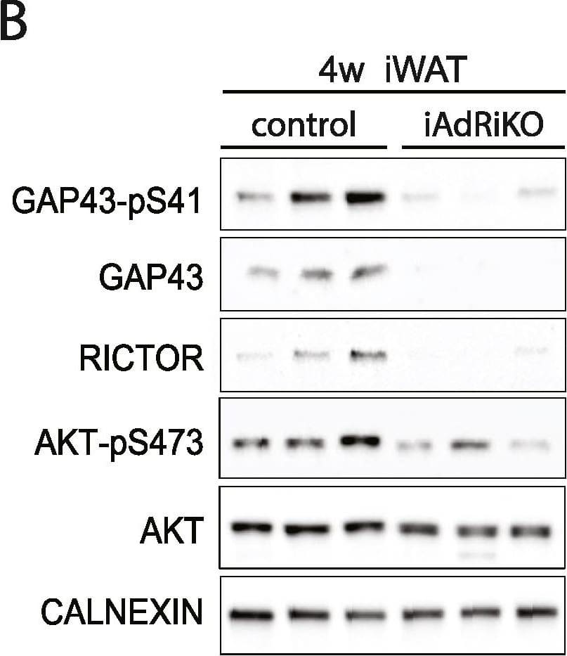
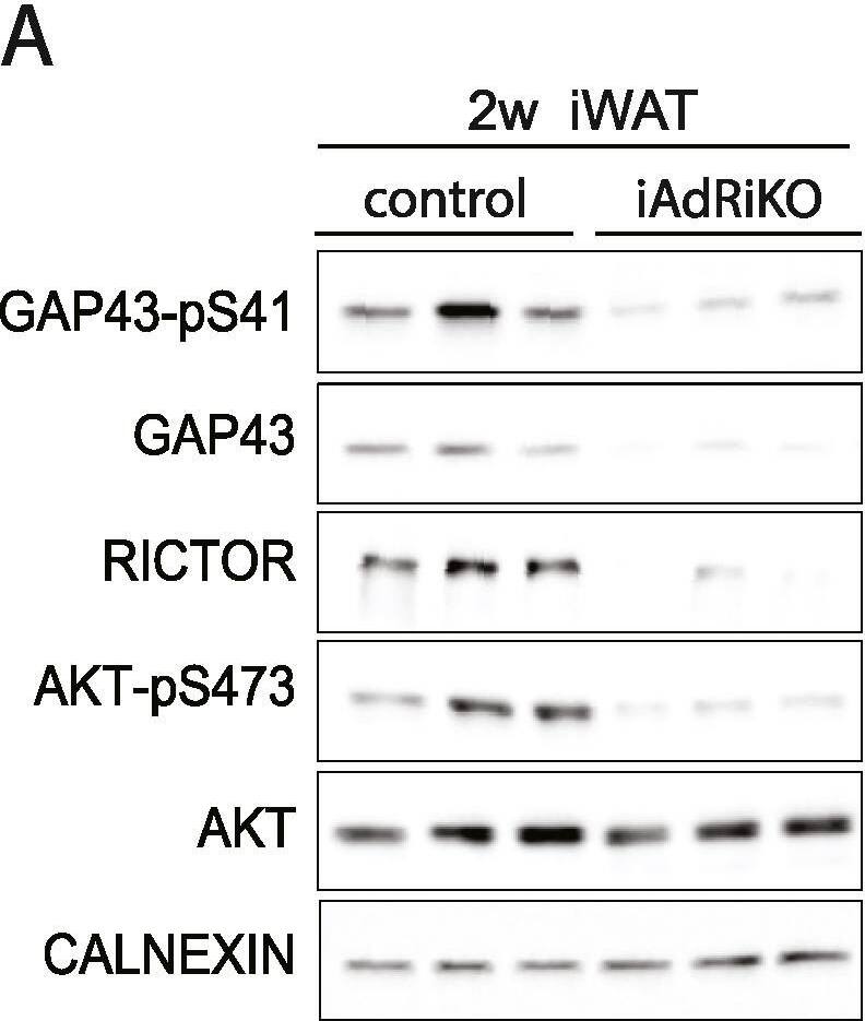
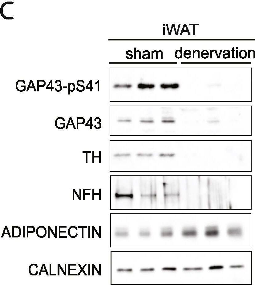
GAP43 expression is downregulated in CGRP-positive neurons upon loss of adipose mTORC2. (A) Immunoblot analysis of inguinal WAT (iWAT) tissue from control and iAdRiKO mice two weeks after tamoxifen treatment. (n = 6; 6). (B) Immunoblot analysis of iWAT tissue from control and iAdRiKO mice four weeks after tamoxifen treatment. (n = 6; 6). (C) Immunoblot analysis of surgically denervated iWAT depot (denervation) compared to iWAT depot from sham-operated mice (sham). Neurofilament heavy polypeptide (NFH). (n = 5; 5). (D) Representative image of a large nerve bundle in iWAT of control mice immunostained with growth-associated protein 43 (GAP43)-pS41 and calcitonin gene-related peptide (CGRP). (N = 11; 9). (E) Representative image of a large nerve bundle in iWAT of control mice immunostained with GAP43-pS41 and tyrosine hydroxylase (TH). (N = 19; 11). Image collected and cropped by CiteAb from the following open publication (https://pubmed.ncbi.nlm.nih.gov/36028121), licensed under a CC-BY license. Not internally tested by R&D Systems.Detection of Mouse GAP-43 by Western Blot
GAP43 expression is downregulated in CGRP-positive neurons upon loss of adipose mTORC2. (A) Immunoblot analysis of inguinal WAT (iWAT) tissue from control and iAdRiKO mice two weeks after tamoxifen treatment. (n = 6; 6). (B) Immunoblot analysis of iWAT tissue from control and iAdRiKO mice four weeks after tamoxifen treatment. (n = 6; 6). (C) Immunoblot analysis of surgically denervated iWAT depot (denervation) compared to iWAT depot from sham-operated mice (sham). Neurofilament heavy polypeptide (NFH). (n = 5; 5). (D) Representative image of a large nerve bundle in iWAT of control mice immunostained with growth-associated protein 43 (GAP43)-pS41 and calcitonin gene-related peptide (CGRP). (N = 11; 9). (E) Representative image of a large nerve bundle in iWAT of control mice immunostained with GAP43-pS41 and tyrosine hydroxylase (TH). (N = 19; 11). Image collected and cropped by CiteAb from the following open publication (https://pubmed.ncbi.nlm.nih.gov/36028121), licensed under a CC-BY license. Not internally tested by R&D Systems.Applications for Phospho-GAP‑43 (S41) Antibody
Application
Recommended Usage
Western Blot
1:1000 dilution
Sample: Rat cortex tissue
Sample: Rat cortex tissue
Formulation, Preparation, and Storage
Purification
Antigen Affinity-purified
Formulation
100 μL in 10 mM HEPES (pH 7.5), 150 mM NaCl, 100 μg/mL BSA and 50% glycerol.
Shipping
The product is shipped with polar packs. Upon receipt, store it immediately at the temperature recommended below.
Stability & Storage
For long-term storage, ≤ -20° C is recommended. Product is stable at ≤ -20° C for at least 1 year.
Background: GAP-43
Long Name
Growth Associated Protein 43
Alternate Names
B-50, Basp2, GAP43, Neuromodulin
Gene Symbol
GAP43
Additional GAP-43 Products
Product Documents for Phospho-GAP‑43 (S41) Antibody
Product Specific Notices for Phospho-GAP‑43 (S41) Antibody
For research use only
Loading...
Loading...
Loading...


