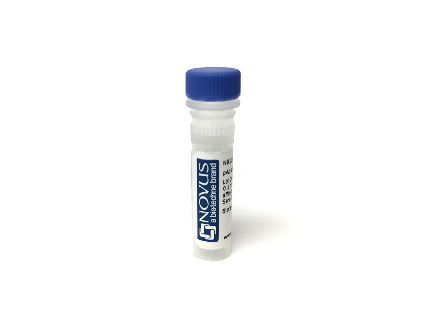
PINK1 Antibody [Janelia Fluor® 669]
Novus Biologicals, part of Bio-Techne | Catalog # NB100-493JF669


Conjugate
Catalog #
Forumulation
Catalog #
Key Product Details
Species Reactivity
Human, Mouse, Rat
Applications
Western Blot
Label
Janelia Fluor 669
Antibody Source
Polyclonal Rabbit IgG
Concentration
Please see the vial label for concentration. If unlisted please contact technical services.
Product Specifications
Immunogen
PINK1 antibody was developed using an N-terminal region synthetic peptide made to the human PINK1 protein sequence (between residues 1-50). [UniProt# Q9BXM7]
Reactivity Notes
Rat (PMID: 24411077) and mouse (PMID: 21760537) reactivity reported in scientific literature
Localization
Mitochondrion
Specificity
PINK1 Antibody is expected recognize isoform 1 but will not recognize isoform 2.
Clonality
Polyclonal
Host
Rabbit
Isotype
IgG
Applications for PINK1 Antibody [Janelia Fluor® 669]
Application
Recommended Usage
Western Blot
Optimal dilutions of this antibody should be experimentally determined.
Application Notes
Optimal dilution of this antibody should be experimentally determined.
Formulation, Preparation, and Storage
Purification
Immunogen affinity purified
Formulation
50mM Sodium Borate
Preservative
0.05% Sodium Azide
Concentration
Please see the vial label for concentration. If unlisted please contact technical services.
Shipping
The product is shipped with polar packs. Upon receipt, store it immediately at the temperature recommended below.
Stability & Storage
Store at 4C in the dark.
Background: PINK1
PINK1 (PTEN induced putative kinase 1) protein contains a N-terminal mitochondrial targeting sequence, putative transmembrane helix, linker region, serine (Ser65)/threonine (Thr257) kinase domain and C-terminal segment. PINK1 is translated in the cytosol, then translocated to the outer mitochondrial membrane where it is rapidly cleaved and degraded as a part of normal mitochondrial function. In damaged (depolarized) mitochondria, PINK1 becomes stabilized and accumulates, resulting in the subsequent phosphorylation of numerous proteins on the mitochondrial surface.
When PINK1 is imported into the cell, mitochondrial processing peptidase, presenilin-associated rhomboid-like protease and AFG3L2 cleave PINK1 and tag it for the ubiquitin-proteasome pathway, keeping low PINK1 protein expression at basal conditions (1,2). Accumulation of PINK1 in mitochondria indicate damage. PINK1 maintains mitochondrial function/integrity, provides protection against mitochondrial dysfunction during cellular stress, and is involved in the clearance of damaged mitochondria via selective autophagy (mitophagy) (3). PINK1 has a theoretical molecular weight of 63 kDa and undergoes proteolytic processing to generate at least two cleaved forms (55 kDa and 42 kDa).
Ultimately PARK2 (E3 Ubiquitin Ligase Parkin) is recruited to the damaged mitochondria where it is activated by 1) PINK-mediated phosphorylation of PARK2 at serine 65, and 2) PARK2 interaction with phosphorylated ubiquitin (also phosphorylated by PINK1 on serine 65) (4,5). There is a strong interplay between Parkin and PINK1, where loss-of-function of human PINK1 results in mitochondrial pathology and can be rescued by Parkin (2,4,5). Mutations in either Parkin or PINK1 alter mitochondrial turnover, resulting in the accumulation of defective mitochondria and, ultimately, neurodegeneration in Parkinson's disease. Mutations in the PINK1 gene located within the PARK6 locus on chromosome 1p35-p36 have been identified in patients with early-onset Parkinson's disease (6).
References
1.Rasool, S., Soya, N., Truong, L., Croteau, N., Lukacs, G. L., & Trempe, J. F. (2018). PINK1 autophosphorylation is required for ubiquitin recognition. EMBO Rep, 19(4). doi:10.15252/embr.201744981
2.Shiba-Fukushima, K., Arano, T., Matsumoto, G., Inoshita, T., Yoshida, S., Ishihama, Y., . . . Imai, Y. (2014). Phosphorylation of mitochondrial polyubiquitin by PINK1 promotes Parkin mitochondrial tethering. PLoS Genet, 10(12), e1004861. doi:10.1371/journal.pgen.1004861
3.Vives-Bauza, C., Zhou, C., Huang, Y., Cui, M., de Vries, R. L., Kim, J., . . . Przedborski, S. (2010). PINK1-dependent recruitment of Parkin to mitochondria in mitophagy. Proc Natl Acad Sci U S A, 107(1), 378-383. doi:10.1073/pnas.0911187107
4.McWilliams, T. G., Barini, E., Pohjolan-Pirhonen, R., Brooks, S. P., Singh, F., Burel, S., . . . Muqit, M. M. K. (2018). Phosphorylation of Parkin at serine 65 is essential for its activation in vivo. Open Biol, 8(11). doi:10.1098/rsob.180108
5.Exner, N., Treske, B., Paquet, D., Holmstrom, K., Schiesling, C., Gispert, S., . . . Haass, C. (2007). Loss-of-function of human PINK1 results in mitochondrial pathology and can be rescued by parkin. J Neurosci, 27(45), 12413-12418. doi:10.1523/jneurosci.0719-07.2007
6.Valente, E. M., Bentivoglio, A. R., Dixon, P. H., Ferraris, A., Ialongo, T., Frontali, M., . . . Wood, N. W. (2001). Localization of a novel locus for autosomal recessive early-onset parkinsonism, PARK6, on human chromosome 1p35-p36. Am J Hum Genet, 68(4), 895-900. doi:10.1086/319522
Long Name
PTEN-induced Putative Kinase 1
Alternate Names
BRPK, PARK6
Gene Symbol
PINK1
Additional PINK1 Products
Product Documents for PINK1 Antibody [Janelia Fluor® 669]
Product Specific Notices for PINK1 Antibody [Janelia Fluor® 669]
Sold under license from the Howard Hughes Medical Institute, Janelia Research Campus.
This product is for research use only and is not approved for use in humans or in clinical diagnosis. Primary Antibodies are guaranteed for 1 year from date of receipt.
Loading...
Loading...
Loading...
Loading...