SARS-CoV-2 Membrane Antibody
R&D Systems, part of Bio-Techne | Catalog # MAB11038

Key Product Details
Species Reactivity
SARS-CoV-2
Applications
Immunocytochemistry, Western Blot
Label
Unconjugated
Antibody Source
Monoclonal Mouse IgG2B Clone # 1046317
Product Specifications
Immunogen
Chinese Hamster Ovary cell line CHO-derived SARS-CoV-2 Membrane (M) protein
Met1-Asn21
Accession # YP_009724393.1
Met1-Asn21
Accession # YP_009724393.1
Specificity
Detects SARS-CoV-2 Membrane (M) protein in direct ELISAs.
Clonality
Monoclonal
Host
Mouse
Isotype
IgG2B
Scientific Data Images for SARS-CoV-2 Membrane Antibody
Detection of SARS-CoV-2 Membrane by Western Blot.
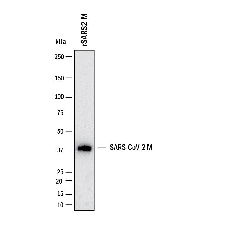
Western blot shows recombinant SARS CoV-2 Membrane. PVDF membrane was probed with 2 µg/mL of Mouse Anti-SARS-CoV-2 Membrane Monoclonal Antibody (Catalog # MAB11038) followed by HRP-conjugated Anti-Mouse IgG Secondary Antibody (HAF018). A specific band was detected for Membrane at approximately 40 kDa (as indicated). This experiment was conducted under reducing conditions and using Western Blot Buffer Group 1.Detection of SARS-CoV-2 Membrane in HEK293 human embryonic kidney cell line transfected.
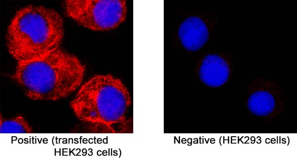
Membrane was detected in immersion fixed HEK293 human embryonic kidney cell line transfected (positive staining) and HEK293 human embryonic kidney cell line (non-transfected, negative staining) using Mouse Anti-SARS-CoV-2 Membrane Monoclonal Antibody (Catalog # MAB11038) at 8 µg/mL for 3 hours at room temperature. Cells were stained using the NorthernLights™ 557-conjugated Anti-Mouse IgG Secondary Antibody (red; Catalog # NL007) and counterstained with DAPI (blue). Specific staining was localized to cytoplasm . View our protocol for Fluorescent ICC Staining of Cells on Coverslips.Applications for SARS-CoV-2 Membrane Antibody
Application
Recommended Usage
Immunocytochemistry
3-25 µg/mL
Sample: immersion fixed HEK293 human embryonic kidney cell line transfected with SARS-CoV-2 Membrane protein
Sample: immersion fixed HEK293 human embryonic kidney cell line transfected with SARS-CoV-2 Membrane protein
Western Blot
2 µg/mL
Sample: Recombinant SARS CoV-2 Membrane protein
Sample: Recombinant SARS CoV-2 Membrane protein
Formulation, Preparation, and Storage
Purification
Protein A or G purified from hybridoma culture supernatant
Reconstitution
Reconstitute at 0.5 mg/mL in sterile PBS. For liquid material, refer to CoA for concentration.
Formulation
Lyophilized from a 0.2 μm filtered solution in PBS with Trehalose. *Small pack size (SP) is supplied either lyophilized or as a 0.2 µm filtered solution in PBS.
Shipping
Lyophilized product is shipped at ambient temperature. Liquid small pack size (-SP) is shipped with polar packs. Upon receipt, store immediately at the temperature recommended below.
Stability & Storage
Use a manual defrost freezer and avoid repeated freeze-thaw cycles.
- 12 months from date of receipt, -20 to -70 °C as supplied.
- 1 month, 2 to 8 °C under sterile conditions after reconstitution.
- 6 months, -20 to -70 °C under sterile conditions after reconstitution.
Background: Membrane
References
- Wu, F. et al. (2020) Nature 579:265.
- Mousavizadeh, L. and S. Ghasemi (2020) J. Microbiol. Immunol. Infect. doi:10.1016/j.jmii.2020.03.022.
- Thomas, S. (2020) Pathog. Immun. 5:342.
- Masters, P.S. (2006) Adv. Virus. Res. 66:193.
- Corse, E. and C.E. Machamer (2003) Virology 312:25.
- Boson, B. et al. (2020) bioRxiv doi:10.1101/2020.08.24.260901.
Long Name
Membrane Glycoprotein
Alternate Names
M protein
Entrez Gene IDs
43740571 (SARS-CoV-2)
Gene Symbol
M
UniProt
Additional Membrane Products
Product Documents for SARS-CoV-2 Membrane Antibody
Product Specific Notices for SARS-CoV-2 Membrane Antibody
For research use only
Loading...
Loading...
Loading...
Loading...

