STAT6 Antibody (177C322.1) - BSA Free
Novus Biologicals, part of Bio-Techne | Catalog # NBP2-25241

![Western Blot: STAT6 Antibody (177C322.1)BSA Free [NBP2-25241] Western Blot: STAT6 Antibody (177C322.1)BSA Free [NBP2-25241]](https://resources.bio-techne.com/images/products/STAT6-Antibody-177C322-1-Western-Blot-NBP2-25241-img0001.jpg)
Conjugate
Catalog #
Forumulation
Catalog #
Key Product Details
Species Reactivity
Validated:
Human, Mouse
Cited:
Human, Mouse
Applications
Validated:
Immunocytochemistry/ Immunofluorescence, Immunoprecipitation, Western Blot
Cited:
Western Blot
Label
Unconjugated
Antibody Source
Monoclonal Mouse IgG1 Clone # 177C322.1
Format
BSA Free
Concentration
1.0 mg/ml
Product Specifications
Immunogen
A portion of amino acids 600-650 of human Signal Transducer and Activator of Transcription 6 was used as the immunogen for the STAT6 Antibody (177C322.1).
Clonality
Monoclonal
Host
Mouse
Isotype
IgG1
Scientific Data Images for STAT6 Antibody (177C322.1) - BSA Free
Western Blot: STAT6 Antibody (177C322.1)BSA Free [NBP2-25241]
Western Blot: STAT6 Antibody (177C322.1) [NBP2-25241] - Analysis of STAT6 in HeLa lysate in the A) absence and B) presence of immunizing peptide using STAT6 antibody at 2 ug/ml. anti mouse Ig HRP secondary antibody and ECL substrate were used for this test.Immunocytochemistry/ Immunofluorescence: STAT6 Antibody (177C322.1) - BSA Free [NBP2-25241]
Immunocytochemistry/Immunofluorescence: STAT6 Antibody (177C322.1) [NBP2-25241] - HeLa cells were fixed for 10 minutes using 10% formalin and then permeabilized for 5 minutes using 1X TBS + 0.5% Triton-X100. The cells were incubated with anti-STAT6 (177C322.1) at 5 ug/ml overnight at 4C and detected with an anti-mouse Dylight 488 (Green) at a 1:500 dilution. Actin was detected with Phalloidin 568 (Red) at a 1:200 dilution. Nuclei were counterstained with DAPI (Blue). Cells were imaged using a 40X objective.STAT6 (177C322.1) in A431 Human Cell Line.
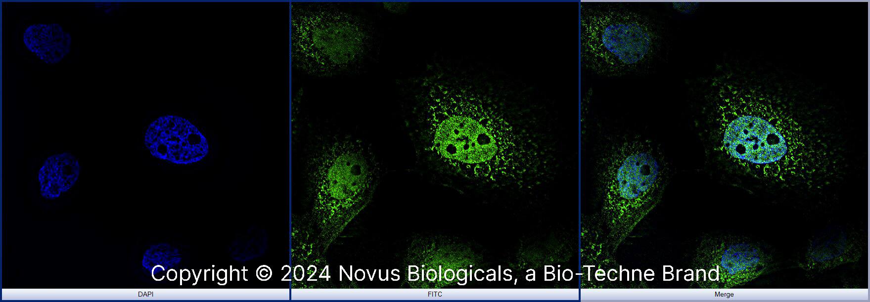
STAT6 (177C322.1) was detected in immersion fixed A431 human skin carcinoma cell line using Mouse anti- STAT6 (177C322.1) Protein-G purified Monoclonal Antibody conjugated to Alexa Fluor® 488 (Catalog # NBP2-25241AF488) (green) at 10 µg/mL overnight at 4C. Cells were counterstained with DAPI (blue). Cells were imaged using a 100X objective and digitally deconvolved.Applications for STAT6 Antibody (177C322.1) - BSA Free
Application
Recommended Usage
Immunocytochemistry/ Immunofluorescence
5 ug/ml
Immunoprecipitation
1-2 ug/ml
Western Blot
1-2 ug/ml
Formulation, Preparation, and Storage
Purification
Protein G purified
Formulation
PBS
Format
BSA Free
Preservative
0.05% Sodium Azide
Concentration
1.0 mg/ml
Shipping
The product is shipped with polar packs. Upon receipt, store it immediately at the temperature recommended below.
Stability & Storage
Store at 4C short term. Aliquot and store at -20C long term. Avoid freeze-thaw cycles.
Background: STAT6
References
1. Waqas, S. F. H., Ampem, G., & Roszer, T. (2019). Analysis of IL-4/STAT6 Signaling in Macrophages. Methods Mol Biol, 1966, 211-224. doi:10.1007/978-1-4939-9195-2_17
2. Goenka, S., & Kaplan, M. H. (2011). Transcriptional regulation by STAT6. Immunol Res, 50(1), 87-96. doi:10.1007/s12026-011-8205-2
Long Name
Signal Transducer and Activator of Transcription 6
Alternate Names
D12S1644, EC 2.4.1.227, EC 2.7.7.6, IL-4 Stat, IL-4-STAT, signal transducer and activator of transcription 6, signal transducer and activator of transcription 6, interleukin-4 induced, STAT, interleukin-4 induced, STAT, interleukin4-induced, STAT6B, STAT6C, transcription factor IL-4 STAT
Gene Symbol
STAT6
UniProt
Additional STAT6 Products
Product Documents for STAT6 Antibody (177C322.1) - BSA Free
Product Specific Notices for STAT6 Antibody (177C322.1) - BSA Free
This product is for research use only and is not approved for use in humans or in clinical diagnosis. Primary Antibodies are guaranteed for 1 year from date of receipt.
Loading...
Loading...
Loading...
Loading...
Loading...
![Immunocytochemistry/ Immunofluorescence: STAT6 Antibody (177C322.1) - BSA Free [NBP2-25241] Immunocytochemistry/ Immunofluorescence: STAT6 Antibody (177C322.1) - BSA Free [NBP2-25241]](https://resources.bio-techne.com/images/products/STAT6-Antibody-177C322-1-Immunocytochemistry-Immunofluorescence-NBP2-25241-img0003.jpg)
