Stathmin-2/STMN2 Antibody - BSA Free Best Seller
Novus Biologicals, part of Bio-Techne | Catalog # NBP1-49461

Conjugate
Catalog #
Key Product Details
Validated by
Knockout/Knockdown, Tagged Protein Expression
Species Reactivity
Validated:
Human, Mouse, Rat, Bovine
Cited:
Human, Mouse, Rat, Avian - Chicken
Applications
Validated:
Immunocytochemistry/ Immunofluorescence, Immunohistochemistry, Immunohistochemistry Whole-Mount, Immunohistochemistry-Frozen, Immunohistochemistry-Paraffin, In vitro assay, In vivo assay, Knockdown Validated, Western Blot
Cited:
IF/IHC, Immunocytochemistry/ Immunofluorescence, Immunohistochemistry, Immunohistochemistry Whole-Mount, Immunohistochemistry-Frozen, Immunohistochemistry-Paraffin, In vivo assay, Western Blot
Label
Unconjugated
Antibody Source
Polyclonal Rabbit IgG
Format
BSA Free
Concentration
1.0 mg/ml
Product Specifications
Immunogen
C-terminal peptide of mouse STMN2. [Swiss-Prot P55821]
Reactivity Notes
Mouse reactivity reported in scientific literature (PMID:32778834).
Clonality
Polyclonal
Host
Rabbit
Isotype
IgG
Theoretical MW
22 kDa.
Disclaimer note: The observed molecular weight of the protein may vary from the listed predicted molecular weight due to post translational modifications, post translation cleavages, relative charges, and other experimental factors.
Disclaimer note: The observed molecular weight of the protein may vary from the listed predicted molecular weight due to post translational modifications, post translation cleavages, relative charges, and other experimental factors.
Scientific Data Images for Stathmin-2/STMN2 Antibody - BSA Free
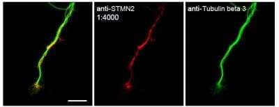
Immunocytochemistry/Immunofluorescence Analysis of Stathmin-2/STMN2 in Mouse DRG Neurons
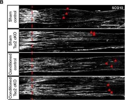
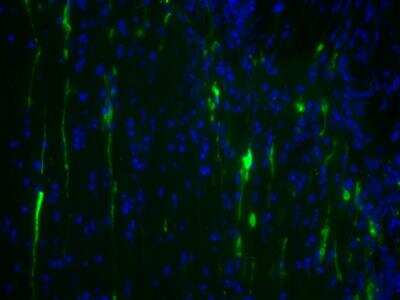
Staining of STMN2 in primary mouse dorsal root ganglia (DRG) neurons Image shows the expected staining of endogenous STMN2 in axons and growth cones. Tubulin is a marker of axons. NBP1-49461 was used at a dilution of 1:4000. Image courtesy of Dr. Jung Eun Shin.Immunohistochemical Staining of Stathmin-2/STMN2 in Frozen Mouse Sciatic Nerves
Stathmin-2-STMN2-Antibody-Immunohistochemistry-Frozen-NBP1-49461-img0015.jpgApplications for Stathmin-2/STMN2 Antibody - BSA Free
Application
Recommended Usage
Immunocytochemistry/ Immunofluorescence
1 - 2 ug/ml
Immunohistochemistry
1:200 - 1:500
Immunohistochemistry Whole-Mount
reported in scientific literature (PMID 35042776)
Immunohistochemistry-Frozen
1:200 - 1:500
Immunohistochemistry-Paraffin
reported in scientific literature (PMID 31182472)
In vitro assay
reported in scientific literature (PMID 22726832)
In vivo assay
reported in scientific literature (PMID 22726832)
Western Blot
1 - 2 ug/ml
Application Notes
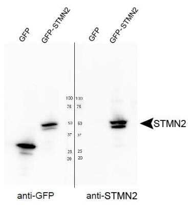
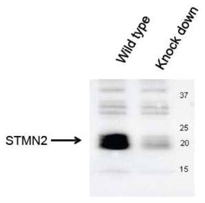
In Western blot a band can be seen at ~22 kDa. The observed molecular weight of the protein may vary from the listed predicted molecular weight due to post translational modifications, post translation cleavages, relative charges, and other experimental factors.
Reviewed Applications
Read 2 reviews rated 4.5 using NBP1-49461 in the following applications:
Formulation, Preparation, and Storage
Purification
Immunogen affinity purified
Formulation
PBS
Format
BSA Free
Preservative
0.02% Sodium Azide
Concentration
1.0 mg/ml
Shipping
The product is shipped with polar packs. Upon receipt, store it immediately at the temperature recommended below.
Stability & Storage
Store at 4C short term. Aliquot and store at -20C long term. Avoid freeze-thaw cycles.
Background: Stathmin-2/STMN2
Alternate Names
SCG10, SCGN10, Stathmin2, STMN2
Gene Symbol
STMN2
Additional Stathmin-2/STMN2 Products
Product Documents for Stathmin-2/STMN2 Antibody - BSA Free
Product Specific Notices for Stathmin-2/STMN2 Antibody - BSA Free
This product is for research use only and is not approved for use in humans or in clinical diagnosis. Primary Antibodies are guaranteed for 1 year from date of receipt.
Loading...
Loading...
Loading...
Loading...
