CellXVivo Mouse Th1 Cell Differentiation Kit
R&D Systems, part of Bio-Techne | Catalog # CDK018

Key Product Details
Assay Procedure
Refer to the product datasheet for complete product details.
Briefly, CD4+ T cells isolated from mouse splenocytes are differentiated ex vivo into the Th1 phenotype with reagents provided in this kit and using the following procedure:
- Isolate CD4+ T cells from mouse splenocytes
- Culture CD4+ T cells in reagents provided in kit
- Verify Th1 cell expansion after 6 days in culture
This kit contains the following reagents for the ex vivo differentiation of mouse Th1 cells.
- Hamster Anti-Mouse CD3, Th1
- Mouse Th1 Reagent 1
- Mouse Th1 Reagent 2
- Mouse Th1 Reagent 3
- Mouse Th1 Reagent 4
- Reconstitution Buffer 1
- Reconstitution Buffer 2
- 20X Wash Buffer
Reagents
- Laboratory mice
- X-VIVO™ 15 Chemically Defined, Serum-free Hematopoietic Cell Medium (Lonza, or equivalent)
- MagCellect™ Mouse Naïve CD4+ T Cell Isolation Kit (R&D Systems, Catalog # MAGM205, or equivalent)
- Penicillin/Streptomycin (optional)
- Cell Activation Cocktail 500X (Tocris®, Catalog # 5476)
Equipment
- Tissue culture plates and/or flasks
- Sterile deionized water
- Pipettes and pipette tips
- Inverted microscope
- Hemocytometer
- 37 °C, 5% CO2 incubator
- Centrifuge
Protocol for mouse Th1 cell differentiation using the CellXVivo™ Mouse Th1 Cell Differentiation Kit.
Note: Results may vary due to strain, age, and/or the health of the mice used for isolation.
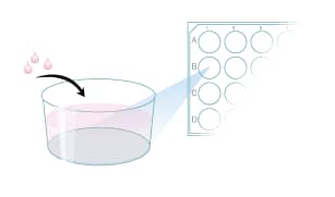
Coat the desired tissue culture plate with Hamster Anti-Mouse CD3,
Th1 antibody.

Isolate mouse splenocytes.

Isolate mouse naïve CD4+ T cells from splenocytes (e.g., using magnetic cell selection).

Perform a cell count.

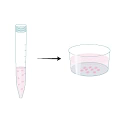
Suspend 1 x 10⁶ naïve CD4+ T cells/mL in Mouse Th1 Differentiation Media. Culture the cells on plates pre-coated with Hamster Anti-Mouse CD3 antibody.

Harvest cells on day 3.
Dilute cells 1:10 with fresh Mouse Th1 Differentiation Media.
Culture cells in a new flask for an additional 3 days.

Verify Th1 cell differentiation by analyzing cytokine expression via flow cytometry or ELISA (optional).


