|
|
Figure 1. Superior expansion of hiPSCs with ExCellerate™ iPSC Expansion Medium.
Human iPSC were maintained in either ExCellerate™ iPSC medium or a competitor’s iPSC medium on different substrates: Recombinant Human Vitronectin (Catalog # 2308-VN) or Cultrex™ UltiMatrix (Catalog # BME001) for 3 days in 6 well plates. Colonies were then dissociated as single cells and counted to obtain fold expansion. Initial cell seeding density was 50,000 cells per well. Graph is displayed as Average ± SEM.
|
|
|
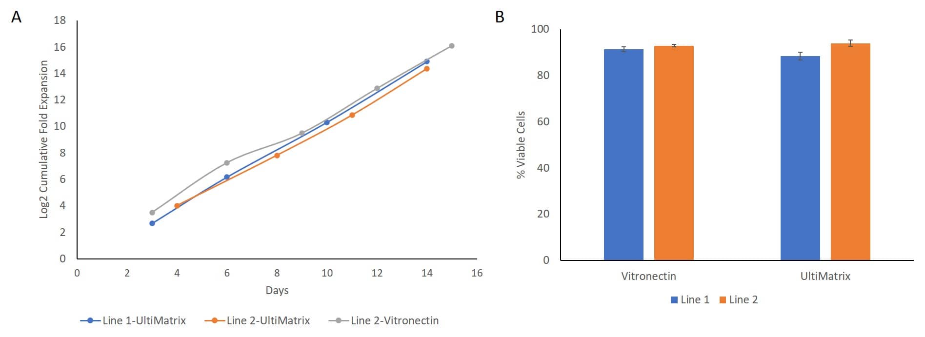
Figure 2. Consistent performance with ExCellerate™ iPSC Expansion Medium across cell lines and matrices.
Different human iPSC lines maintained in ExCellerate™ iPSC Expansion Medium showed minimal line-to-line variability in cumulative fold expansion over 15 days (A) and exhibit greater than ~90% viability after 4 days in culture (B). Graph is displayed as average ± standard deviation. Cell lines were cultured on Cultrex™ UltiMatrix (Catalog # BME001), Recombinant Human Vitronectin (Catalog # 2308-VN).
|
|
|
Figure 3. Human iPSCs cultured in ExCellerate™ iPSC Expansion Medium show characteristics of classic hiPSC colony morphology consistently across cell lines.
Different hiPSC lines grown using Cultrex UltiMatrix (Catalog # BME001) and cultured in ExCellerate™ iPSC Expansion Medium show healthy rounded colony morphology and characteristics such as compact cells and clearly defined smooth colony edges.
|
|
|
Figure 4. HiPSCs cultured in ExCellerate™ iPSC Expansion Medium maintain expression of stemness markers over long-term culture.
HiPSC lines grown using Cultrex UltiMatrix (Catalog # BME001) and maintained in ExCellerate™ iPSC Expansion Medium express undifferentiated stem cell markers OCT3/4 (red, Catalog # AF1759) and Tra-1-60 (red, Catalog # MAB4770) along with F-actin (green) and DAPI (blue, Catalog # 5748) (A). HiPSC lines express high levels of OCT3/4, SSEA-4, SOX2, and no SSEA-1 as assessed by flow cytometry (Catalog # FMC001). Undifferentiated stem cell marker expression is greater than 97% across 4 cell lines after more than 45 passages (B-C). Graph is displayed as average ± standard deviation.
|
|
|
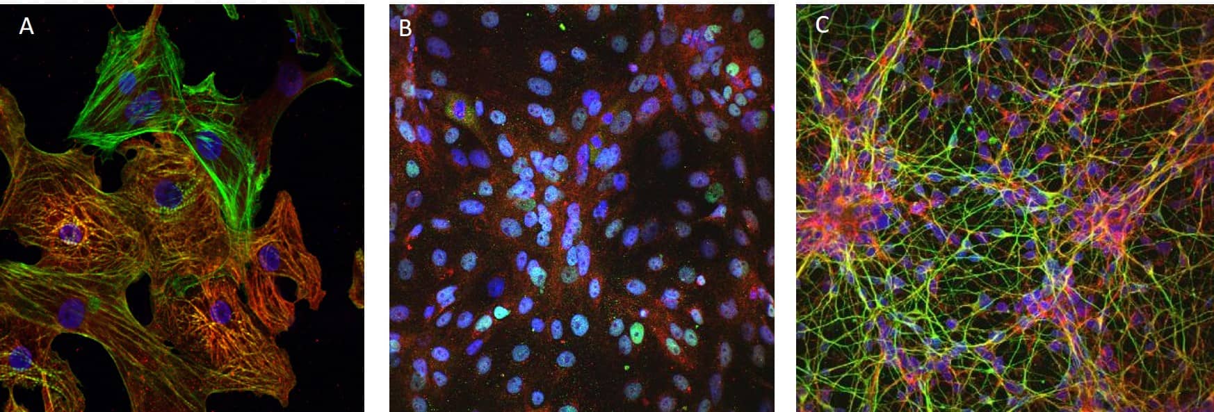
Figure 5. HiPSCs maintained in ExCellerate™ iPSC Expansion Medium differentiate into cardiomyocytes, hepatocytes and neurons.
HiPSCs maintain their pluripotency in ExCellerate™ iPSC Expansion Medium and can differentiate into cells of the three germ layers (Catalog # SC027B). Shown here are images of hiPSC derived cardiomyocytes (mesoderm, Catalog # SC032B) expressing cardiac troponin T (cTNT1, red, Catalog # MAB1874), F-actin (green) and DAPI (blue, Catalog # 5748) (A); hepatocytes (endoderm, Catalog # SC033) showing expression of albumin (red, Catalog # MAB1455), HNF4a (green, Catalog # PP-H1415) and DAPI (blue) (B); and neurons (ectoderm, Catalog # SC035) expressing N-cadherin (red), Tuj1 (green) and DAPI (blue)(C). Cells were grown using Cultrex UltiMatrix (Catalog # BME001).
|
|
|
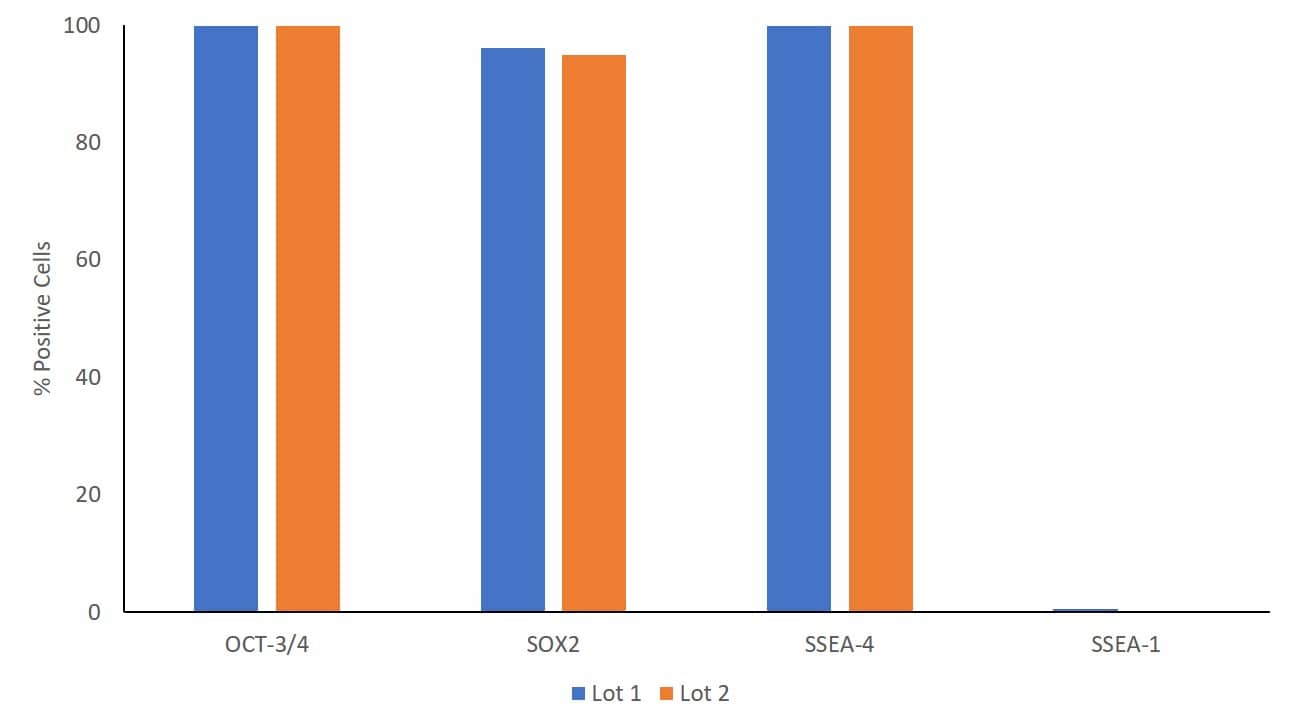
Figure 6. Lot-to-lot consistency with ExCellerate™ iPSC Expansion Medium.
Human iPSC lines grown using Cultrex UltiMatrix (Catalog # BME001) and cultured in different lots of ExCellerate™ iPSC Expansion Medium express high levels of undifferentiated stem cell markers OCT-3/4, SOX2, SSEA-4 and no SSEA-1 as assessed flow cytometry (Catalog # FMC001).
|
|
|
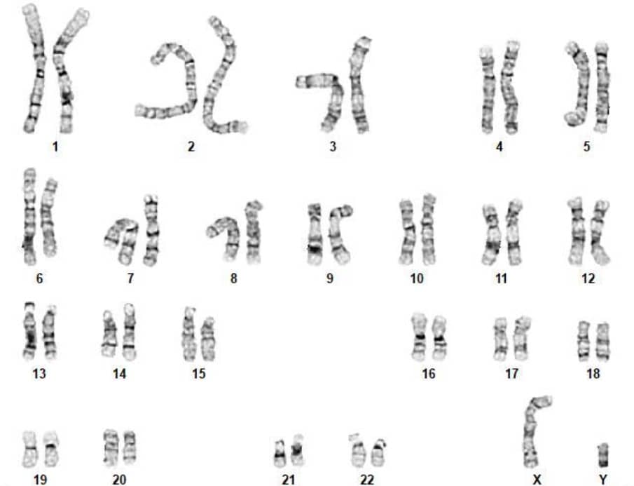
Figure 7. HiPSCs cultured in ExCellerate™ iPSC Expansion Medium show normal karyotype.
HiPSC lines grown using Cultrex UltiMatrix (Catalog # BME001) and maintained in ExCellerate™ iPSC Expansion Medium for more than 30 passages display normal karyotype.
|


