Recombinant Human B7-H3 His-tag Avi-tag Protein, CF
R&D Systems, part of Bio-Techne | Catalog # AVI2318

Key Product Details
Source
Accession #
Structure / Form
Conjugate
Applications
Product Specifications
Source
| Human B7-H3 (Leu29-Thr461) Accession # Q5ZPR3-1 |
HHHHHH | Avi-tag |
| N-terminus | C-terminus |
Purity
Endotoxin Level
N-terminal Sequence Analysis
Predicted Molecular Mass
SDS-PAGE
Activity
The ED50 for this effect is 3-12 μg/mL.
Measured by its binding ability in a functional ELISA.
When Human B7-H3 Antibody (Catalog # AF1027) is immobilized at 1.0 μg/mL, 100 μL/well, the concentration of Biotinylated Recombinant Human B7‑H3 His-tag Avi-tag (Catalog # AVI2318) that produces 50% of the optimal binding response is approximately 3 -15 ng/mL.
Scientific Data Images for Recombinant Human B7-H3 His-tag Avi-tag Protein, CF
Recombinant Human B7-H3 His-tag Avi-tag Protein Binding Activity
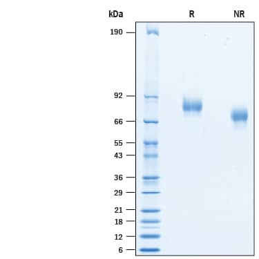
When Human B7-H3 Antibody (Catalog # AF1027) is immobilized at 1.0 µg/mL, 100 µL/well, the concentration of Biotinylated Recombinant Human B7-H3 His-tag Avi-tag (Catalog # AVI2318) that produces 50% of the optimal binding response is approximately 3-15 ng/mL.Recombinant Human B7-H3 His-tag Avi-tag Protein SDS-PAGE
2 μg/lane of Recombinant Human B7-H3 His-tag Avi-tag (Catalog # AVI2318) was resolved with SDS-PAGE under reducing (R) and non-reducing (NR) conditions and visualized by Coomassie® Blue staining, showing bands at 70-90 kDa.Formulation, Preparation and Storage
AVI2318
| Formulation | Lyophilized from a 0.2 μm filtered solution in PBS with Trehalose. |
| Reconstitution | Reconstitute at 200 μg/mL in PBS. |
| Shipping | The product is shipped at ambient temperature. Upon receipt, store it immediately at the temperature recommended below. |
| Stability & Storage | Use a manual defrost freezer and avoid repeated freeze-thaw cycles.
|
Background: B7-H3
Human B7 homolog 3 (B7-H3) is a member of the B7 family of immune proteins that provide signals for the regulation of immune responses (1 - 3). Other family members include B7-1, B7-2, B7-H1/PD-L1, B7-H2, and PD-L2. B7 family proteins are type I transmembrane immunoglobulin (Ig) superfamily members that contain extracellular Ig V‑like and Ig C‑like domains with a short cytoplasmic tail. Among the family members there is about 20 - 40% amino acid (aa) sequence identity. B7-H3 was initially reported to be a 316 aa type I transmembrane precursor protein that contained a signal sequence, an extracellular region with one V‑type and one C‑type Ig domain, a transmembrane segment and a short cytoplasmic tail (1). Subsequent studies have identified a second 110 kDa form whose precursor is 534 aa in length. Termed 4IgB7-H3 or B7-H3b, this molecule has two additional Ig-like domains (one V‑type and one C‑type) and shows a ubiquituous expression pattern (4, 5). It would appear that the human 4Ig form is the principal, if not the only form of B7-H3 (5). Its precursor contains a 26 aa signal sequence, a 435 aa extracellular region, a 31 aa transmembrane domain, and a 42 aa cytoplasmic tail. The four Ig-like domains alternate between V‑type and C‑type, and apparently are the consequence of a V‑C type tandem duplication (4, 5). B7-H3b is expressed on dendritic cells as well as activated T, B and NK cells (5). The mouse gene differs from that of human in that it cannot code for four Ig-like domains; only a V‑type:C‑type pair (4). Human B7-H3b binding to an undefined receptor has shown to be inhibitory to NK cells and cytokine release (6). It also seems to be required for late stage osteoblast differentiation (7).
References
- Chapoval, A.I. et al. (2001) Nat. Immunol. 2:269.
- Sharpe, A.H. and G.J. Freeman (2002) Nat. Rev. Immunol. 2:116.
- Coyle, A. and J.Gutierrez-Ramos (2001) Nat. Immunol. 2:203.
- Sun, M. et al. (2002) J. Immunol. 168:6294.
- Steinberger, P. et al. (2004) J. Immunol. 172:2352.
- Prasad, D.V.R. et al. (2004) J. Immunol. 173:2500.
- Suh, W-K. et al. (2004) Proc. Natl. Acad. Sci. USA 101:12969.
Long Name
Alternate Names
Gene Symbol
UniProt
Additional B7-H3 Products
Product Documents for Recombinant Human B7-H3 His-tag Avi-tag Protein, CF
Product Specific Notices for Recombinant Human B7-H3 His-tag Avi-tag Protein, CF
For research use only

