Recombinant Mouse R-Spondin 2 Fc Chimera Protein, CF
R&D Systems, part of Bio-Techne | Catalog # 11567-RS

Key Product Details
Source
Structure / Form
Conjugate
Applications
Product Specifications
Source
| Mouse RSPO-2 (Ala32-Gly205) Accession # Q8BFU0.1 |
IEGRMD | Human IgG1 (Pro100-Lys330) |
| N-terminus | C-terminus |
Purity
Endotoxin Level
N-terminal Sequence Analysis
Predicted Molecular Mass
SDS-PAGE
Activity
Scientific Data Images for Recombinant Mouse R-Spondin 2 Fc Chimera Protein, CF
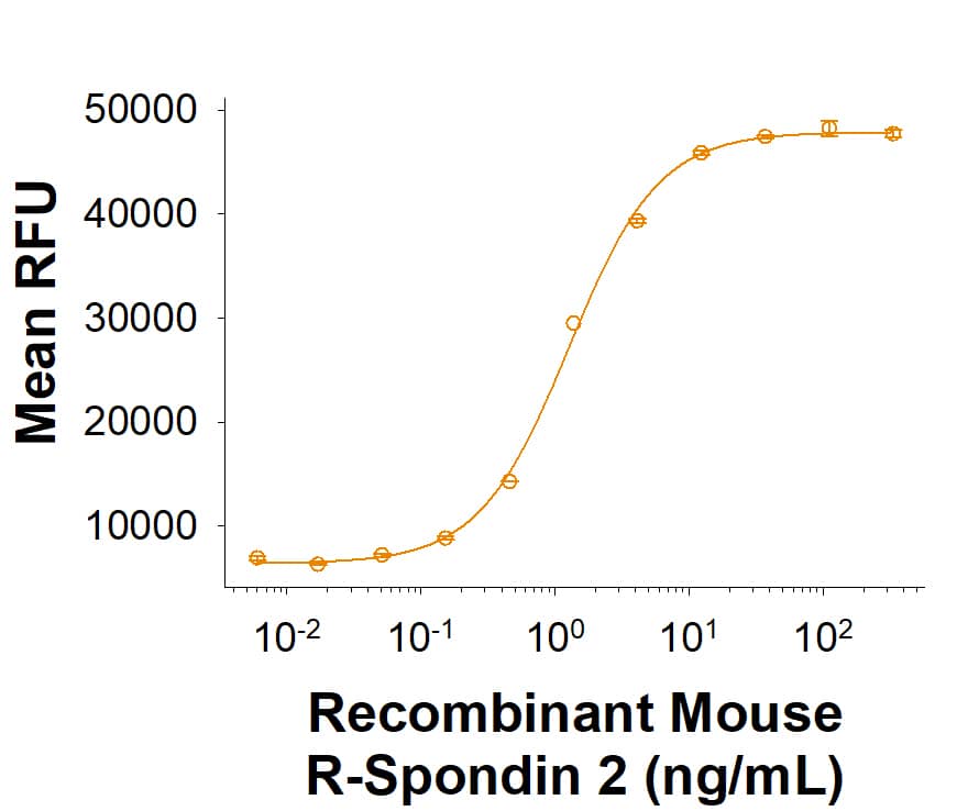
Recombinant Mouse R‑Spondin 2 Fc Chimera Protein Bioactivity.
Recombinant Mouse R-Spondin 2 Fc Chimera Protein (Catalog # 11567-RS) activates TCF reporter activity in HEK293 human embryonic kidney cells in the presence of Wnt-3a. The ED50 for this effect is 0.600-12.0 ng/mL.Recombinant Mouse R‑Spondin 2 Fc Chimera Protein SDS-PAGE.
2 μg/lane of Recombinant Mouse R‑Spondin 2 Fc Chimera Protein (Catalog # 11567-RS) was resolved with SDS-PAGE under reducing (R) and non-reducing (NR) conditions and visualized by Coomassie® Blue staining, showing bands at 52-63 kDa and 104-130 kDa, respectively.Formulation, Preparation and Storage
11567-RS
| Formulation | Lyophilized from a 0.2 μm filtered solution in PBS with Trehalose. |
| Reconstitution | Reconstitute the 20 μg size at 200 μg/mL in PBS. Reconstitute all other sizes at 500 μg/mL in PBS. |
| Shipping | The product is shipped at ambient temperature. Upon receipt, store it immediately at the temperature recommended below. |
| Stability & Storage | Use a manual defrost freezer and avoid repeated freeze-thaw cycles.
|
Background: R-Spondin 2
Roof plate-specific Spondin 2 isoform 1 (R‑Spondin 2, RSPO2), also known as cysteine‑rich and single thrombospondin domain containing protein 2 (Cristin 2), is a 33 kDa secreted protein that belongs to the R‑Spondin family (1‑3). The four R‑Spondins regulate Wnt/ beta-catenin signaling and overlap in expression and function (1‑3). Like other R‑Spondins, RSPO2 contains two adjacent cysteine‑rich furin-like domains (aa 90‑134) followed by a thrombospondin (TSP-1) motif (aa 144‑204) and a C-terminal region rich in basic residues (aa 207‑243). The basic region binds heparin and mediates cell surface retention and extracellular matrix attachment while the furin-like domains are required for Wnt/ beta-catenin signaling (1, 3, 4). RSPO2 contains one potential N‑glycosylation site. Mature mouse RSPO2 shares 97‑98% aa identity with human, rat, equine, canine and bovine RSPO2 and ~40% aa identity with RSPO1, RSPO3 and RSPO4. One potential 237 aa mouse isoform diverges after aa 206 and lacks a basic region (5). Human RSPO2 is expressed in organs of endodermal origin in adults, including intestine and lung, and is down‑regulated in tumors of these tissues (1). In the embryonic mouse, RSPO2 expression is concentrated in the apical epidermal ridge, hippocampus, and developing muscle, teeth and bones (1, 6). Deletion of RSPO2 results in down‑regulation of Wnt activity in these areas, malformations of the facial skeleton and limbs, and respiratory failure at birth (7-9). RSPO2 is an extracellular potentiator of Wnt/ beta-catenin signaling (3, 4). It functions at least in part by binding LRP-6, stimulating its long-term phosphorylation and down‑regulating its internalization (3, 4). RSPO proteins, especially RSPO2 and RSPO3, also antagonize DKK1 activity by interfering with DKK1‑mediated LRP-6 and Kremen association (10).
References
- Kazanskaya, O. et al. (2004) Dev. Cell 7:525.
- Kim, K.-A. et al. (2006) Cell Cycle 5:23.
- Nam, J.-S. et al. (2006) J. Biol. Chem. 281:13247.
- Li, S-J. et al. (2009) Cell Signal. 21:916.
- Entrez accession # EDL08755.
- Nam, J.-S. et al. (2007) Gene Expr. Patterns 7:306.
- Yamada, W. et al. (2009) Biochem. Biophys. Res. Commun. 381:453.
- Jin, Y.-R. et al. (2011) Dev. Biol. 352:1.
- Nam, J.-S. et al. (2007) Dev. Biol. 311:124.
- Kim, K.-A. et al. (2007) Mol. Biol. Cell 19:2588.
Long Name
Alternate Names
Gene Symbol
Additional R-Spondin 2 Products
Product Documents for Recombinant Mouse R-Spondin 2 Fc Chimera Protein, CF
Product Specific Notices for Recombinant Mouse R-Spondin 2 Fc Chimera Protein, CF
For research use only

