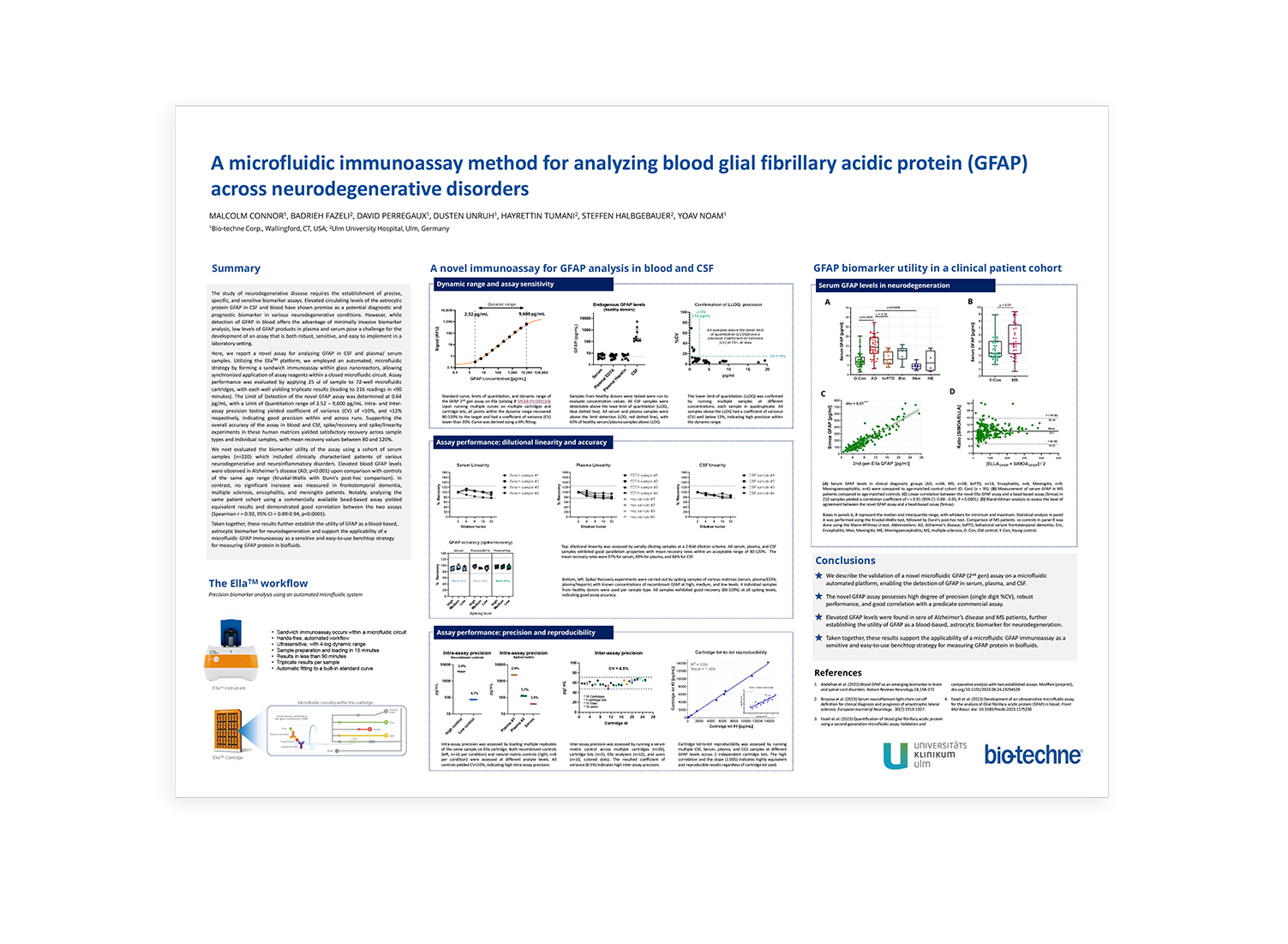
A Microfluidic Strategy for the Analysis of Blood Glial Fibrillary Acidic Protein (GFAP) Across Neurodegenerative Disorders
Scientific Meeting PostersThe study of neurodegenerative disease requires the establishment of precise, specific, and sensitive biomarker assays. Elevated circulating levels of astrocytic protein GFAP in CSF and blood have shown promise as a potential diagnostic and prognostic biomarker in various neurodegenerative conditions. However, while detection of GFAP in blood offers the advantage of minimally invasive biomarker analysis, low levels of GFAP products in plasma and serum pose a challenge for the development of an assay that is both robust and sensivite and easy to implement in a laboratory setting.
Here, we report a novel assay for analyzing GFAP in CSF and plasma/serum samples. Utilizing the EllaTM platform, we employed an automated, microfluidic strategy by forming a sandwich immunoassay within glass nanoreactors, allowing synchronized application of assay reagents within a closed microfluidic circuit. Assay performance was evaluated by applying 25 uL of sample to 72-well microfluidic cartridges, with each well yielding triplicate results (leading to 216 readings in <90 minutes). The Limit of Detection of the novel GFAP assay was determined at 0.64 pg/mL, with a Limit of Quantitation range of 2.5 – 9,600 pg/mL. Intra- and inter- assay precision testing yielded a coefficient of variance (CV) of <10%, and <12% respectively, indicating good precision within and across runs. Supporting the overall accuracy of the assay in blood and CSF, spike/recovery and spike/linearity experiments in these human matrices yielded satisfactory recovery across sample types and individual samples, with mean recovery values between 80 and 120%. We next evaluated the biomarker utility of the assay using a cohort of serum samples (n=220) which included clinically characterized patients of various neurodegenerative and neuroinflammatory disorders. Elevated blood GFAP levels were observed in Alzheimer’s disease (AD; p<0.001) upon comparison of controls from the same age range (Kruskal-Wallis with Dunn’s post-hoc comparison). In contrast, no significant increase was measured in frontotemporal dementia, multiple sclerosis, encephalitis, and meningitis patients. Notable, analyzing the sample patient cohort using a commercially available bead-based assay yielded equivalent results and demonstrated good correlation between the two assays (Spearman r = 0.92, 95%CI = 0.89-0.94, p<0.001). Taken together, these results further establish the utility of GFAP as a blood-based astrocytic biomarker for neurodegeneration and support the applicability of a microfluidic GFAP immunoassay as a sensitive and easy-to-use benchtop strategy for measuring GFAP protein in biofluids.
