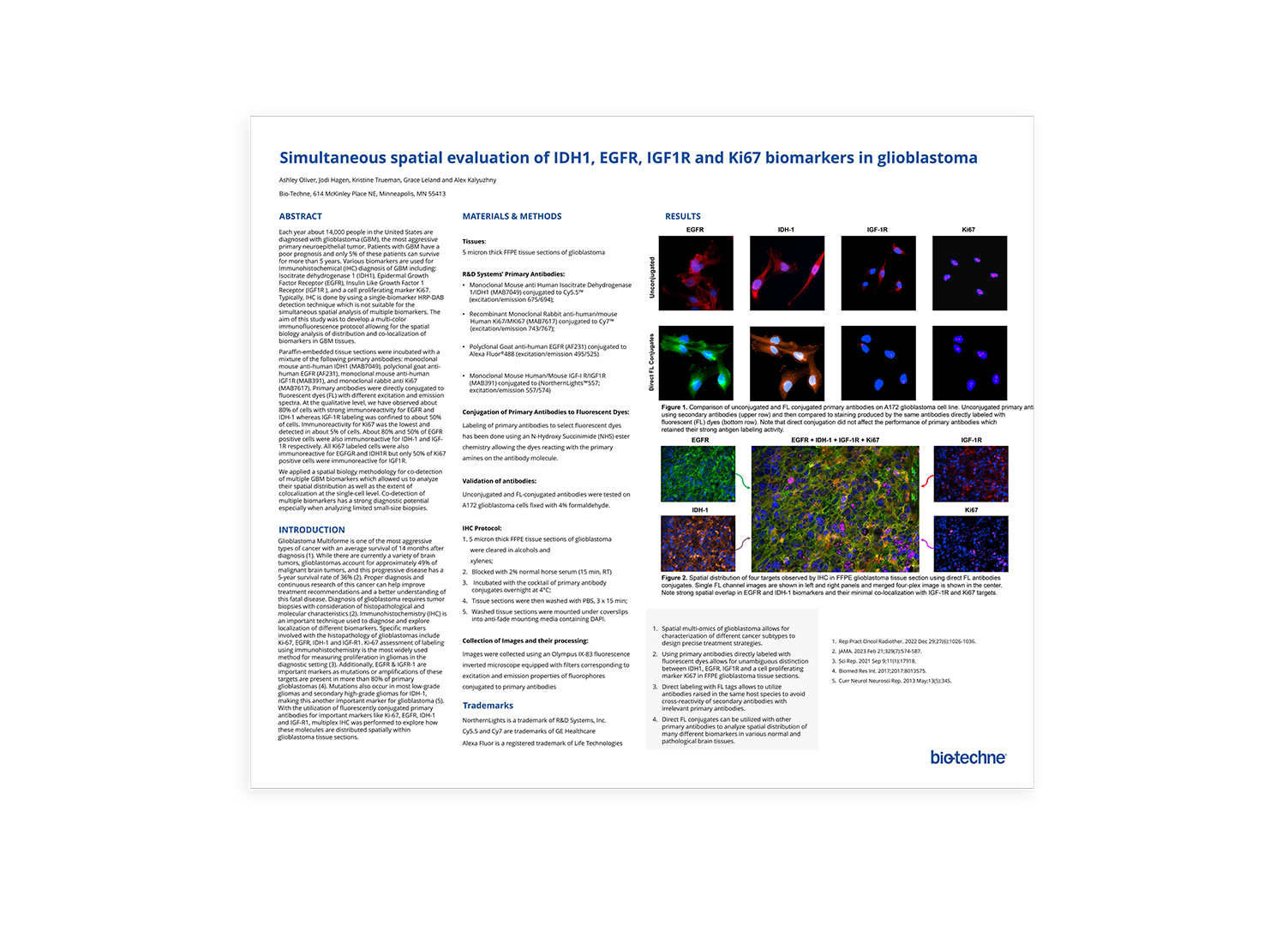
Simultaneous Spatial Evaluation of IDH1, EGFR, IGF1R and Ki67 Biomarkers in Glioblastoma
Scientific Meeting PostersEach year about 14,000 people in the United States are diagnosed with glioblastoma (GBM), the most aggressive primary neuroepithelial tumor. Patients with GBM have a poor prognosis and only 5% of these patients can survive for more than 5 years. Various biomarkers are used for Immunohistochemical (IHC) diagnosis of GBM including: Isocitrate dehydrogenase 1 (IDH1), Epidermal Growth Factor Receptor (EGFR), Insulin Like Growth Factor 1 Receptor (IGF1R), and a cell proliferating marker Ki67. Typically, IHC is done by using a single-biomarker HRP-DAB detection technique which is not suitable for the simultaneous spatial analysis of multiple biomarkers.
The aim of this study was to develop a multi-color immunofluorescence protocol allowing for the spatial biology analysis of distribution and co-localization of biomarkers in GBM tissues. Paraffin-embedded tissue sections were incubated with a mixture of the following primary antibodies: monoclonal mouse anti-human IDH1 (MAB7049), polyclonal goat anti-human EGFR (AF231), monoclonal mouse anti-human IGF1R (MAB391), and monoclonal rabbit anti Ki67 (MAB7617). For detection, we used species specific secondary antibodies conjugated to fluorescent dyes with different excitation and emission spectra. At the qualitative level, we have observed about 80% of cells with strong immunoreactivity for EGFR and IDH-1 whereas IGF-1R labeling was confined to about 50% of cells. Immunoreactivity for Ki67 was the lowest and detected in about 5% of cells. About 80% and 50% of EGFR positive cells were also immunoreactive for IDH-1 and IGF-1R respectively. All Ki67 labeled cells were also immunoreactive for EGFGR and IDH1R but only 50% of Ki67 positive cells were immunoreactive for IGF1R. We applied a spatial biology methodology for co-detection of multiple GBM biomarkers which allowed us to analyze their spatial distribution as well as the extent of colocalization at the single-cell level. Co-detection of multiple biomarkers has a strong diagnostic potential especially when analyzing limited small-size biopsies.
