Dr. Li Yu
"Work on things that are most interesting to you and pay much less attention to whether these things are important or not. "
I’m Li Yu, and this is why I research.
Dr Li Yu is a Faculty Member in the School of Life Sciences, Tsinghua University. He trained as a postdoctoral fellow at the National Institutes of Health (NIH), USA. During his time at the laboratory of Dr. Michael Leonardo, M.D., Dr. Li Yu discovered autophagic lysosome reformation. As a faculty member at the School of Life Sciences, Tsinghua University, Beijing, Dr. Li Yu continued his research on the molecular mechanism regulating autophagy and autophagic lysosome reformation. More recently however his research has shifted towards a new direction following his discovery of a new cellular organelle which forms vesicle structures identifiable in the retraction fibers of migrating cells. They have coined the name “migrasomes” for this novel organelle.
Would you tell us about your career path and what first sparked your interest in science?
I had a typical career path, got my PhD in Beijing University, did a postdoc at the NIH, and then joined Tsinghua as a faculty member. If there is anything special, it is that I was a really bad undergraduate student. I think I was very close to the bottom of our class, if not at the bottom myself. Ironically, my interest in research stems from my undergraduate thesis work, so I learned first-hand how serendipity works in research.

Your lab has made significant discoveries in autophagy regulation and function. Which discovery in this area has been more exciting to you?
My work on autophagic lysosome reformation excites me the most, because we discovered something new rather than addressing a long-standing question. Personally, I like this type of work.
With the discovery of migrasomes by your group, what are some exciting questions being investigated?
We are currently trying to answer the following questions about migrasomes: What genes regulate migrasome formation? How are migrasomes formed? What are the physiological functions of migrasomes? Do migrasomes play any roles in diseases?
Discovering a new organelle must have been thrilling. Could you tell us about the initial observation and the main experiments that solidified this finding?
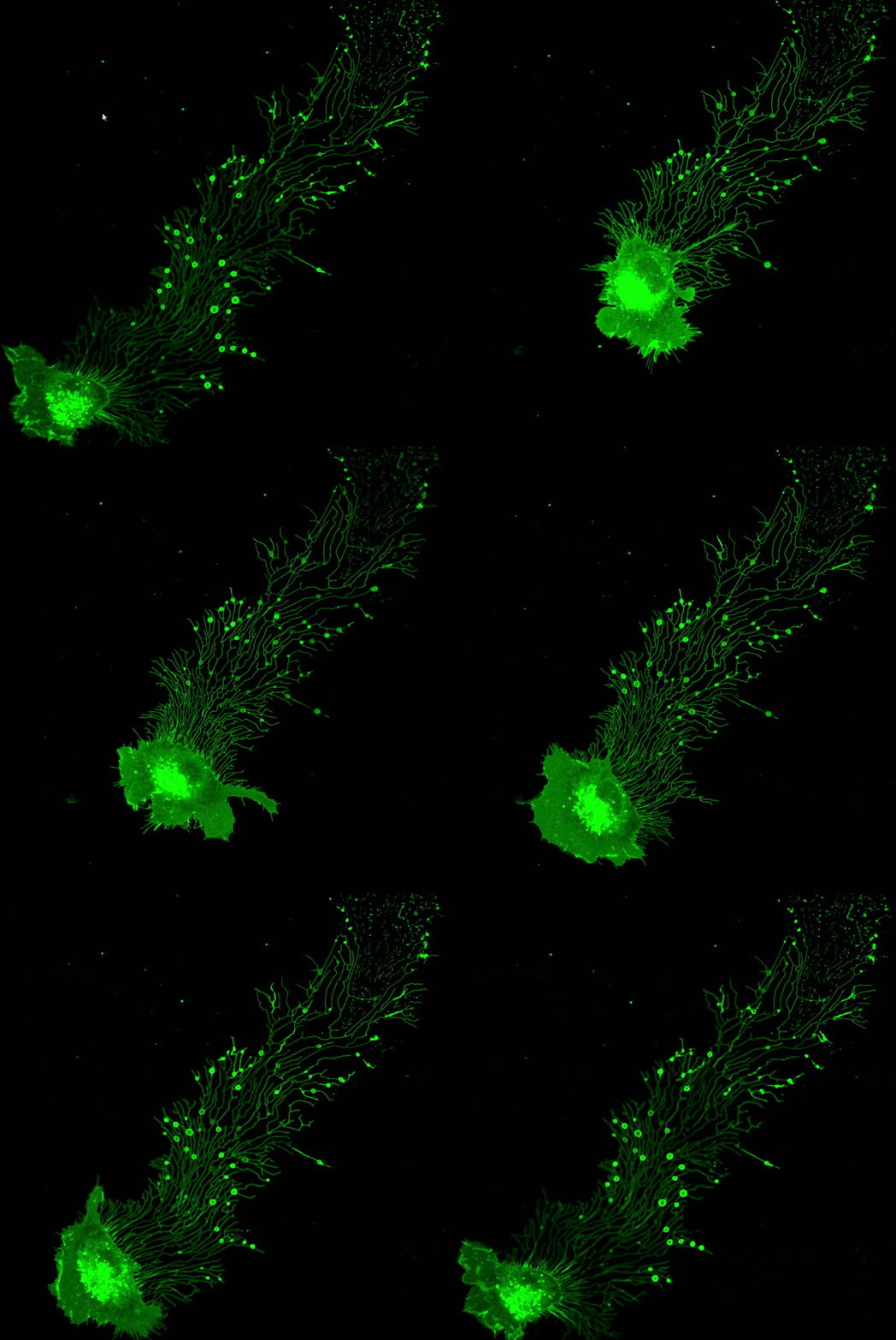
In 2010, I first observed large extracellular vesicles with luminal vesicles when I collected transmission electron microscope (TEM) images for an autophagy-related project. By the way, I do a lot of TEM myself, and I am still doing it today. After I noticed these structures, I started to look for them intentionally when I did TEM. Very soon I realized these structures are not freaks, and I decided I needed to work on them. For a modern-day cell biologist, the first thing you do when you find an unknown structure is to find a marker for optical microscopy observations. To do that, we purified migrasomes by fractionation and then we used mass spectroscopy to identify the proteins enriched on migrasomes. Very soon we found that Tspan4 is highly enriched on migrasomes. Using Tspan4-GFP as a marker, we quickly observed migrasome formation using live-cell imaging. With that data, I knew I had found something new.

Migrasomes in Action! Migracytosis is the migration-dependent release of cytosolic content through migrasomes. Courtesy of Dr. Li Yu’s Lab
Migrasomes and exosomes are both types of extracellular vesicles. How do they compare in form and function?
First, we don’t think that migrasomes are extracellular vesicles. Detached migrasomes are a type of extracellular vesicle for sure; however, we have evidence to show that many functions of migrasomes are carried out before migrasomes are detached from the cell body. That’s why we think that migrasomes are organelles rather than a type of extracellular vesicle. Generating extracellular vesicles (detached migrasomes) is just one of many functions of migrasomes.
Detached migrasomes are a type of extracellular vesicle, but the differences between detached migrasomes and exosomes are huge. I’ll list just a few examples here. The size is different: exosomes are 50-60 nm, while migrasomes are 1-3 µm. The protein composition is vastly different: there is only a 20% overlap between migrasomes and exosomes. They are regulated by different genetic pathways and the biogenesis process is completely different: exosomes are first generated as luminal vesicles for multiple vesicular bodies (MVBs), and exosomes are released when MVBs fuse with the plasma membrane; in contrast, migrasomes are formed by assembly of macrodomains on the plasma membrane.
Although we are just beginning to investigate the function of migrasomes, our recent work shows that there are key differences between migrasomes and exosomes. Migrasomes are enriched with signaling molecules such as chemokines, cytokine and growth factors, and migrasomes carry out their functions by sending signals to other cells. I am not aware that sending signaling ligands to other cells has been reported in exosomes.
One more thing to add, migrasomes mark the track of the migrating cell which generates them, and thus they encode spatial information. I think this is one of the key features which could have very important physiological implications.
Your discovery of migrasomes has propelled you towards a completely new research area. What are some challenges and benefits of embarking in a new research focus?
The most difficult part is developing technology and reagents to study migrasomes. Migrasomes are new, so we had to develop everything from scratch. Take for example the in vivo imaging of migrasomes – since migrasomes are small in size, the popular systems for in vivo imaging do not work very well. Fortunately, we are able to work with an excellent team to build a microscope for this purpose.
As for the benefits, there are a lot. Here are a few examples: no competition yet, we can do the work at a pace we like; we can enjoy the freedom of a new field and do whatever we like; by working on something new, it is easy to get students excited…..
What is your advice for young scientists interested in pursuing a research career?
Work on things that are most interesting to you and pay much less attention to whether these things are important or not. Often, you can never know whether a new thing is important until your work reaches a certain point.
And finally, if you could meet any scientist from the past, who would you meet and why?
George Gamow, he was very creative and never afraid to venture into new things. Most importantly, he was super fun, and he always took science as a diversion and made conferences into parties.