Chikungunya Virus Products
Chikungunya virus (CHIKV) belongs to the family of alphaviruses and is transmitted by Aedes mosquitoes resulting in infection characterized by symptoms including arthritis-like joint pain and inflammation, fever, rash, and headache (1-3). The CHIKV genome consists of an 11.8 kilobase (kb) positive-sense, single-stranded RNA virus that has two open reading frames (ORFs) (1-3). The first ORF at the 5' end encodes a 2472 amino acid (aa) nonstructural polyprotein and the ORF at the 3' end encodes a 1244 aa structural polyprotein (1-3). The polyproteins generate four nonstructural proteins (nsP1-4) and five structural proteins: capsid (C), envelope E1, E2, E3, and 6K (1-3). The nonstructural proteins comprise CHIKV's RNA replicase (1,2). Structurally, CHIKV has a diameter of ~65 nm and the virion is formed by an icosahedral nucleocapsid shell encapsulating genomic RNA and surrounded by a lipid bilayer envelope (2,3).
Although analysis suggests CHIKV originated in Africa over 500 years ago, first infections weren't reported until the 1950s (1-3). CHIKV has evolved three distinct genotypes, or strains, based on location, termed West African (WA), East/Central/Southern African (ECSA), and Asian (2-3). The WA strain has been most closely associated with enzootic transmission whereas the ECSA strain contributes more to urban epidemics (2). Nonhuman primates are the primary reservoir for the viral host with transmission occurring via mosquitos biting and infecting humans (1-3). Upon acute infection, the virus replicates in cells including fibroblasts and macrophages resulting in innate immune response in infected tissues characterized by infiltrating cells like macrophages, monocytes, natural killer (NK) cells, and lymphocytes (2,3). Infection results in increased production of proinflammatory cytokines such as interferon alpha (IFN-alpha) and interleukin 6 (IL-6), chemokines, and growth factors (2,3). Physical manifestations of infection are high fever, polyarthralgia, headache, arthritis, and rash (1-3). CHIKV symptoms can be confused with other infections like those from dengue fever and Zika virus (1-3). There are no specific antivirals or vaccines for CHIKV, but rather symptoms are treated with antipyretics and non-steroidal anti-inflammatory drugs (NSAIDs) (2,3). While in vitro culture models and in vivo rodent and non-human primate models have been used to study CHIKV pathogenesis and advance our knowledge of the disease, the specific cellular mechanisms are not fully understood (3).
References
1. Vu DM, Jungkind D, Angelle Desiree LaBeaud. Chikungunya Virus. Clin Lab Med. 2017;37(2):371-382. https://doi.org/10.1016/j.cll.2017.01.008
2. Silva LA, Dermody TS. Chikungunya virus: epidemiology, replication, disease mechanisms, and prospective intervention strategies. J Clin Invest. 2017;127(3):737-749. https://doi.org/10.1172/JCI84417
3. Ganesan VK, Duan B, Reid SP. Chikungunya Virus: Pathophysiology, Mechanism, and Modeling. Viruses. 2017;9(12):368. Published 2017 Dec 1. https://doi.org/10.3390/v9120368
Show More
Although analysis suggests CHIKV originated in Africa over 500 years ago, first infections weren't reported until the 1950s (1-3). CHIKV has evolved three distinct genotypes, or strains, based on location, termed West African (WA), East/Central/Southern African (ECSA), and Asian (2-3). The WA strain has been most closely associated with enzootic transmission whereas the ECSA strain contributes more to urban epidemics (2). Nonhuman primates are the primary reservoir for the viral host with transmission occurring via mosquitos biting and infecting humans (1-3). Upon acute infection, the virus replicates in cells including fibroblasts and macrophages resulting in innate immune response in infected tissues characterized by infiltrating cells like macrophages, monocytes, natural killer (NK) cells, and lymphocytes (2,3). Infection results in increased production of proinflammatory cytokines such as interferon alpha (IFN-alpha) and interleukin 6 (IL-6), chemokines, and growth factors (2,3). Physical manifestations of infection are high fever, polyarthralgia, headache, arthritis, and rash (1-3). CHIKV symptoms can be confused with other infections like those from dengue fever and Zika virus (1-3). There are no specific antivirals or vaccines for CHIKV, but rather symptoms are treated with antipyretics and non-steroidal anti-inflammatory drugs (NSAIDs) (2,3). While in vitro culture models and in vivo rodent and non-human primate models have been used to study CHIKV pathogenesis and advance our knowledge of the disease, the specific cellular mechanisms are not fully understood (3).
References
1. Vu DM, Jungkind D, Angelle Desiree LaBeaud. Chikungunya Virus. Clin Lab Med. 2017;37(2):371-382. https://doi.org/10.1016/j.cll.2017.01.008
2. Silva LA, Dermody TS. Chikungunya virus: epidemiology, replication, disease mechanisms, and prospective intervention strategies. J Clin Invest. 2017;127(3):737-749. https://doi.org/10.1172/JCI84417
3. Ganesan VK, Duan B, Reid SP. Chikungunya Virus: Pathophysiology, Mechanism, and Modeling. Viruses. 2017;9(12):368. Published 2017 Dec 1. https://doi.org/10.3390/v9120368
36 results for "Chikungunya Virus" in Products
36 results for "Chikungunya Virus" in Products
Chikungunya Virus Products
Chikungunya virus (CHIKV) belongs to the family of alphaviruses and is transmitted by Aedes mosquitoes resulting in infection characterized by symptoms including arthritis-like joint pain and inflammation, fever, rash, and headache (1-3). The CHIKV genome consists of an 11.8 kilobase (kb) positive-sense, single-stranded RNA virus that has two open reading frames (ORFs) (1-3). The first ORF at the 5' end encodes a 2472 amino acid (aa) nonstructural polyprotein and the ORF at the 3' end encodes a 1244 aa structural polyprotein (1-3). The polyproteins generate four nonstructural proteins (nsP1-4) and five structural proteins: capsid (C), envelope E1, E2, E3, and 6K (1-3). The nonstructural proteins comprise CHIKV's RNA replicase (1,2). Structurally, CHIKV has a diameter of ~65 nm and the virion is formed by an icosahedral nucleocapsid shell encapsulating genomic RNA and surrounded by a lipid bilayer envelope (2,3).
Although analysis suggests CHIKV originated in Africa over 500 years ago, first infections weren't reported until the 1950s (1-3). CHIKV has evolved three distinct genotypes, or strains, based on location, termed West African (WA), East/Central/Southern African (ECSA), and Asian (2-3). The WA strain has been most closely associated with enzootic transmission whereas the ECSA strain contributes more to urban epidemics (2). Nonhuman primates are the primary reservoir for the viral host with transmission occurring via mosquitos biting and infecting humans (1-3). Upon acute infection, the virus replicates in cells including fibroblasts and macrophages resulting in innate immune response in infected tissues characterized by infiltrating cells like macrophages, monocytes, natural killer (NK) cells, and lymphocytes (2,3). Infection results in increased production of proinflammatory cytokines such as interferon alpha (IFN-alpha) and interleukin 6 (IL-6), chemokines, and growth factors (2,3). Physical manifestations of infection are high fever, polyarthralgia, headache, arthritis, and rash (1-3). CHIKV symptoms can be confused with other infections like those from dengue fever and Zika virus (1-3). There are no specific antivirals or vaccines for CHIKV, but rather symptoms are treated with antipyretics and non-steroidal anti-inflammatory drugs (NSAIDs) (2,3). While in vitro culture models and in vivo rodent and non-human primate models have been used to study CHIKV pathogenesis and advance our knowledge of the disease, the specific cellular mechanisms are not fully understood (3).
References
1. Vu DM, Jungkind D, Angelle Desiree LaBeaud. Chikungunya Virus. Clin Lab Med. 2017;37(2):371-382. https://doi.org/10.1016/j.cll.2017.01.008
2. Silva LA, Dermody TS. Chikungunya virus: epidemiology, replication, disease mechanisms, and prospective intervention strategies. J Clin Invest. 2017;127(3):737-749. https://doi.org/10.1172/JCI84417
3. Ganesan VK, Duan B, Reid SP. Chikungunya Virus: Pathophysiology, Mechanism, and Modeling. Viruses. 2017;9(12):368. Published 2017 Dec 1. https://doi.org/10.3390/v9120368
Show More
Although analysis suggests CHIKV originated in Africa over 500 years ago, first infections weren't reported until the 1950s (1-3). CHIKV has evolved three distinct genotypes, or strains, based on location, termed West African (WA), East/Central/Southern African (ECSA), and Asian (2-3). The WA strain has been most closely associated with enzootic transmission whereas the ECSA strain contributes more to urban epidemics (2). Nonhuman primates are the primary reservoir for the viral host with transmission occurring via mosquitos biting and infecting humans (1-3). Upon acute infection, the virus replicates in cells including fibroblasts and macrophages resulting in innate immune response in infected tissues characterized by infiltrating cells like macrophages, monocytes, natural killer (NK) cells, and lymphocytes (2,3). Infection results in increased production of proinflammatory cytokines such as interferon alpha (IFN-alpha) and interleukin 6 (IL-6), chemokines, and growth factors (2,3). Physical manifestations of infection are high fever, polyarthralgia, headache, arthritis, and rash (1-3). CHIKV symptoms can be confused with other infections like those from dengue fever and Zika virus (1-3). There are no specific antivirals or vaccines for CHIKV, but rather symptoms are treated with antipyretics and non-steroidal anti-inflammatory drugs (NSAIDs) (2,3). While in vitro culture models and in vivo rodent and non-human primate models have been used to study CHIKV pathogenesis and advance our knowledge of the disease, the specific cellular mechanisms are not fully understood (3).
References
1. Vu DM, Jungkind D, Angelle Desiree LaBeaud. Chikungunya Virus. Clin Lab Med. 2017;37(2):371-382. https://doi.org/10.1016/j.cll.2017.01.008
2. Silva LA, Dermody TS. Chikungunya virus: epidemiology, replication, disease mechanisms, and prospective intervention strategies. J Clin Invest. 2017;127(3):737-749. https://doi.org/10.1172/JCI84417
3. Ganesan VK, Duan B, Reid SP. Chikungunya Virus: Pathophysiology, Mechanism, and Modeling. Viruses. 2017;9(12):368. Published 2017 Dec 1. https://doi.org/10.3390/v9120368
Applications: ICC/IF, B/N, ChIP
Reactivity:
Virus
| Reactivity: | Virus |
| Details: | Human IgG1 kappa Monoclonal Clone #E26D9.02 |
| Applications: | ICC/IF, B/N, ChIP |
Applications: IHC, WB, ELISA, ICC/IF, Func
Reactivity:
Virus
| Reactivity: | Virus |
| Details: | Mouse IgM Monoclonal Clone #3E7b |
| Applications: | IHC, WB, ELISA, ICC/IF, Func |
Applications: IHC, WB, ELISA, ICC/IF, Func
Reactivity:
Virus
| Reactivity: | Virus |
| Details: | Mouse IgM Monoclonal Clone #8A2c |
| Applications: | IHC, WB, ELISA, ICC/IF, Func |
Applications: ICC/IF, B/N, ChIP
Reactivity:
Virus
| Reactivity: | Virus |
| Details: | Human IgG1 kappa Monoclonal Clone #E26G8 |
| Applications: | ICC/IF, B/N, ChIP |
Applications: ICC/IF, B/N, ChIP
Reactivity:
Virus
| Reactivity: | Virus |
| Details: | Human IgG1 Monoclonal Clone #E04C1.02 |
| Applications: | ICC/IF, B/N, ChIP |
Applications: WB, ICC/IF, Flow
Reactivity:
Virus
| Reactivity: | Virus |
| Details: | Mouse IgG2B Monoclonal Clone #6A11 |
| Applications: | WB, ICC/IF, Flow |
Applications: WB, ICC/IF, Flow
Reactivity:
Virus
| Reactivity: | Virus |
| Details: | Mouse IgG2B Monoclonal Clone #6A11 |
| Applications: | WB, ICC/IF, Flow |
Applications: WB, ICC/IF, Flow
Reactivity:
Virus
| Reactivity: | Virus |
| Details: | Mouse IgG2B Monoclonal Clone #6A11 |
| Applications: | WB, ICC/IF, Flow |
Applications: WB, ICC/IF, Flow
Reactivity:
Virus
| Reactivity: | Virus |
| Details: | Mouse IgG2B Monoclonal Clone #6A11 |
| Applications: | WB, ICC/IF, Flow |
Applications: WB, ICC/IF, Flow
Reactivity:
Virus
| Reactivity: | Virus |
| Details: | Mouse IgG2B Monoclonal Clone #6A11 |
| Applications: | WB, ICC/IF, Flow |
Applications: WB, ICC/IF, Flow
Reactivity:
Virus
| Reactivity: | Virus |
| Details: | Mouse IgG2B Monoclonal Clone #6A11 |
| Applications: | WB, ICC/IF, Flow |
Applications: WB, ICC/IF, Flow
Reactivity:
Virus
| Reactivity: | Virus |
| Details: | Mouse IgG2B Monoclonal Clone #6A11 |
| Applications: | WB, ICC/IF, Flow |
Applications: WB, ICC/IF, Flow
Reactivity:
Virus
| Reactivity: | Virus |
| Details: | Mouse IgG2B Monoclonal Clone #6A11 |
| Applications: | WB, ICC/IF, Flow |
Applications: WB, ICC/IF, Flow
Reactivity:
Virus
| Reactivity: | Virus |
| Details: | Mouse IgG2B Monoclonal Clone #6A11 |
| Applications: | WB, ICC/IF, Flow |
Applications: WB, ICC/IF, Flow
Reactivity:
Virus
| Reactivity: | Virus |
| Details: | Mouse IgG2B Monoclonal Clone #6A11 |
| Applications: | WB, ICC/IF, Flow |
Applications: WB, ICC/IF, Flow
Reactivity:
Virus
| Reactivity: | Virus |
| Details: | Mouse IgG2B Monoclonal Clone #6A11 |
| Applications: | WB, ICC/IF, Flow |
Applications: WB, ICC/IF, Flow
Reactivity:
Virus
| Reactivity: | Virus |
| Details: | Mouse IgG2B Monoclonal Clone #6A11 |
| Applications: | WB, ICC/IF, Flow |
Applications: WB, ICC/IF, Flow
Reactivity:
Virus
| Reactivity: | Virus |
| Details: | Mouse IgG2B Monoclonal Clone #6A11 |
| Applications: | WB, ICC/IF, Flow |
Applications: WB, ICC/IF, Flow
Reactivity:
Virus
| Reactivity: | Virus |
| Details: | Mouse IgG2B Monoclonal Clone #6A11 |
| Applications: | WB, ICC/IF, Flow |
Applications: WB, ICC/IF, Flow
Reactivity:
Virus
| Reactivity: | Virus |
| Details: | Mouse IgG2B Monoclonal Clone #6A11 |
| Applications: | WB, ICC/IF, Flow |
Applications: WB, ICC/IF, Flow
Reactivity:
Virus
| Reactivity: | Virus |
| Details: | Mouse IgG2B Monoclonal Clone #6A11 |
| Applications: | WB, ICC/IF, Flow |
Applications: WB, ICC/IF, Flow
Reactivity:
Virus
| Reactivity: | Virus |
| Details: | Mouse IgG2B Monoclonal Clone #6A11 |
| Applications: | WB, ICC/IF, Flow |
Applications: WB, ICC/IF, Flow
Reactivity:
Virus
| Reactivity: | Virus |
| Details: | Mouse IgG2B Monoclonal Clone #6A11 |
| Applications: | WB, ICC/IF, Flow |
Applications: WB, ICC/IF, Flow
Reactivity:
Virus
| Reactivity: | Virus |
| Details: | Mouse IgG2B Monoclonal Clone #6A11 |
| Applications: | WB, ICC/IF, Flow |
Applications: WB, ICC/IF, Flow
Reactivity:
Virus
| Reactivity: | Virus |
| Details: | Mouse IgG2B Monoclonal Clone #6A11 |
| Applications: | WB, ICC/IF, Flow |


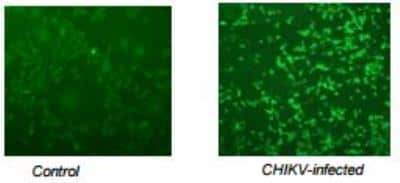
![Immunocytochemistry/ Immunofluorescence: Chikungunya Virus Antibody (3E7b) [NBP2-53118] Immunocytochemistry/ Immunofluorescence: Chikungunya Virus Antibody (3E7b) [NBP2-53118]](https://resources.bio-techne.com/images/products/Chikungunya-virus-Antibody-3E7b-Immunocytochemistry-Immunofluorescence-NBP2-53118-img0001.jpg)
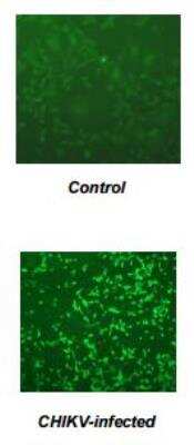
![Immunocytochemistry/ Immunofluorescence: Chikungunya Virus Antibody (8A2c) [NBP2-53117] Immunocytochemistry/ Immunofluorescence: Chikungunya Virus Antibody (8A2c) [NBP2-53117]](https://resources.bio-techne.com/images/products/Chikungunya-virus-Antibody-8A2c-Immunocytochemistry-Immunofluorescence-NBP2-53117-img0001.jpg)


![Chikungunya Virus Antibody (6A11) [mFluor Violet 450 SE] [NBP2-53111MFV450] -](https://resources.bio-techne.com/images/products/nbp2-53111mfv450_mouse-chikungunya-virus-mab-6a11-mfluor-violet-450-se-226202312582326.png)
![Chikungunya Virus Antibody (6A11) [Alexa Fluor® 350] [NBP2-53111AF350] - Chikungunya Virus Antibody (6A11) [Alexa Fluor® 350]](https://resources.bio-techne.com/images/products/nbp2-53111af350_mouse-chikungunya-virus-mab-6a11-alexa-fluor-350-29820239331766.jpg)
![Chikungunya Virus Antibody (6A11) [mFluor Violet 500 SE] -](https://resources.bio-techne.com/images/products/nbp2-53111mfv500_mouse-chikungunya-virus-mab-6a11-mfluor-violet-500-se-2762023952297.png)