Western Blot: beta Tubulin AntibodyBSA Free [NB600-936]
Western Blot: beta Tubulin Antibody - BSA Free [NB600-936] - beta Tubulin Antibody [NB600-936] - Reduced levels of NADPH oxidases 1 and 4 at 18% versus 5% O2. (a) Representative Western blots showing Nox1 and Beta-tubulin in PC-3 cells or Nox4 and Beta-tubulin in C2C12 cells, at 5% and 18% O2. (b, c) Average Nox1 signal How Supraphysiological Oxygen Levels in Standard Cell Culture Affect Oxygen-Consuming Reactions. Image collected and cropped by CiteAb from the following publication (//pubmed.ncbi.nlm.nih.gov/30363917/) licensed under a CC-BY license.
Immunocytochemistry/ Immunofluorescence: beta Tubulin Antibody - BSA Free [NB600-936]
Immunocytochemistry/Immunofluorescence: beta Tubulin Antibody [NB600-936] - PC12 cells were fixed in 4% paraformaldehyde for 10 minutes and permeabilized in 0.05% Triton X-100 in PBS for 5 minutes. The cells were incubated with anti-beta Tubulin Antibody (NB600-936) at 2 ug/ml overnight at 4C and detected with an anti-rabbit Dylight 488 (Green) at a 1:1000 dilution for 60 minutes. Nuclei were counterstained with DAPI (Blue). Cells were imaged using a 100X objective and digitally deconvolved.
Immunohistochemistry-Paraffin: beta Tubulin Antibody - BSA Free [NB600-936]
Immunohistochemistry-Paraffin: beta Tubulin Antibody [NB600-936] - Analysis of FFPE tissue section of normal human skin using 1:2000 dilution of beta Tubulin antibody. Intense cytoplasmic staining of beta Tubulin (TUBB) protein was observed in various cells of the epidermal as well as the dermal cells [10X Magnification].
Western Blot: beta Tubulin AntibodyBSA Free [NB600-936]
Western Blot: beta Tubulin Antibody [NB600-936] - Analysis in HeLa whole cell lysate at a 1:1,000 dilution.
Western Blot: beta Tubulin AntibodyBSA Free [NB600-936]
Western Blot: beta Tubulin Antibody [NB600-936] - Expression of pluripotency related markers and a sphere culture of BEAS-2B under normoxia. HIF-2alpha expression was detected in the nucleus of BEAS-2B under normoxia by western blotting. Beta tubulin and TATA binding protein (TBP) were used as protein markers for the cytosol and nucleus fraction, respectively. Image collected and cropped by CiteAb from the following publication (https://www.nature.com/articles/srep29311), licensed under a CC-BY license.
Immunocytochemistry/ Immunofluorescence: beta Tubulin Antibody - BSA Free [NB600-936]
Immunocytochemistry/Immunofluorescence: beta Tubulin Antibody [NB600-936] - Confocal immunofluorescent analysis of C2C12 cells using beta Tubulin antibody (NB600-936, 1:5). An Alexa Fluor 488-conjugated Goat to rabbit IgG was used as secondary antibody (green). Actin filaments were labeled with Alexa Fluor 568 phalloidin (red). DAPI was used to stain the cell nuclei (blue).
Immunocytochemistry/ Immunofluorescence: beta Tubulin Antibody - BSA Free [NB600-936]
Immunocytochemistry/Immunofluorescence: beta Tubulin Antibody [NB600-936] - Analysis of beta Tubulin in mouse hippocampal primary culture. Image courtesy of product review by Lin Yi-Wen.
Immunocytochemistry/ Immunofluorescence: beta Tubulin Antibody - BSA Free [NB600-936]
Immunocytochemistry/Immunofluorescence: beta Tubulin Antibody [NB600-936] - NIH3T3 cells were fixed in 4% paraformaldehyde for 10 minutes and permeabilized in 0.05% Triton X-100 in PBS for 5 minutes. The cells were incubated with anti-beta Tubulin Antibody (NB600-936) at 2 ug/ml overnight at 4C and detected with an anti-rabbit Dylight 488 (Green) at a 1:1000 dilution for 60 minutes. Nuclei were counterstained with DAPI (Blue). Cells were imaged using a 100X objective and digitally deconvolved.
Immunohistochemistry-Paraffin: beta Tubulin Antibody - BSA Free [NB600-936]
Immunohistochemistry-Paraffin: beta Tubulin Antibody [NB600-936] - Analysis of FFPE tissue section of human esophageal squamous cell carcinoma (SCC) using 1:2000 dilution of beta Tubulin antibody. Strong cytoplasmic immuno-positivity of beta Tubulin (TUBB) was observed in SCC cells as well as the associated tumor stromal cells [10X Magnification].
Immunohistochemistry-Paraffin: beta Tubulin Antibody - BSA Free [NB600-936]
Immunohistochemistry-Paraffin: beta Tubulin Antibody [NB600-936] - Analysis of FFPE tissue section of human esophageal squamous cell carcinoma (SCC) using 1:2000 dilution of beta Tubulin antibody. This representative image shows a cytoplasm specific staining of beta Tubulin (TUBB) in SCC cells [60X Magnification].
Immunohistochemistry-Paraffin: beta Tubulin Antibody - BSA Free [NB600-936]
Immunohistochemistry-Paraffin: beta Tubulin Antibody [NB600-936] - Analysis of FFPE tissue section of normal human brain using 1:2000 dilution of beta Tubulin antibody. The various brain cells depicted strong cytoplasmic immunoreactivity of beta Tubulin (TUBB) protein [10X Magnification].
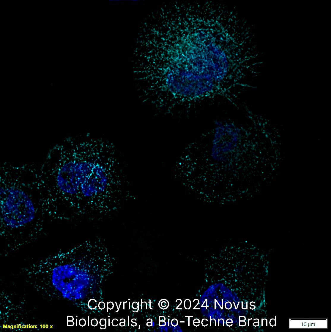
Beta Tubulin in A431 Human Cell Line.
Beta Tubulin was detected in immersion fixed A431 human skin carcinoma cell line using Rabbit anti-beta Tubulin Affinity Purified Polyclonal Antibody conjugated to Alexa Fluor® 647 (Catalog # NB600-936AF647) (light blue) at 5 µg/mL overnight at 4C. Cells were counterstained with DAPI (blue). Cells were imaged using a 100X objective and digitally deconvolved.
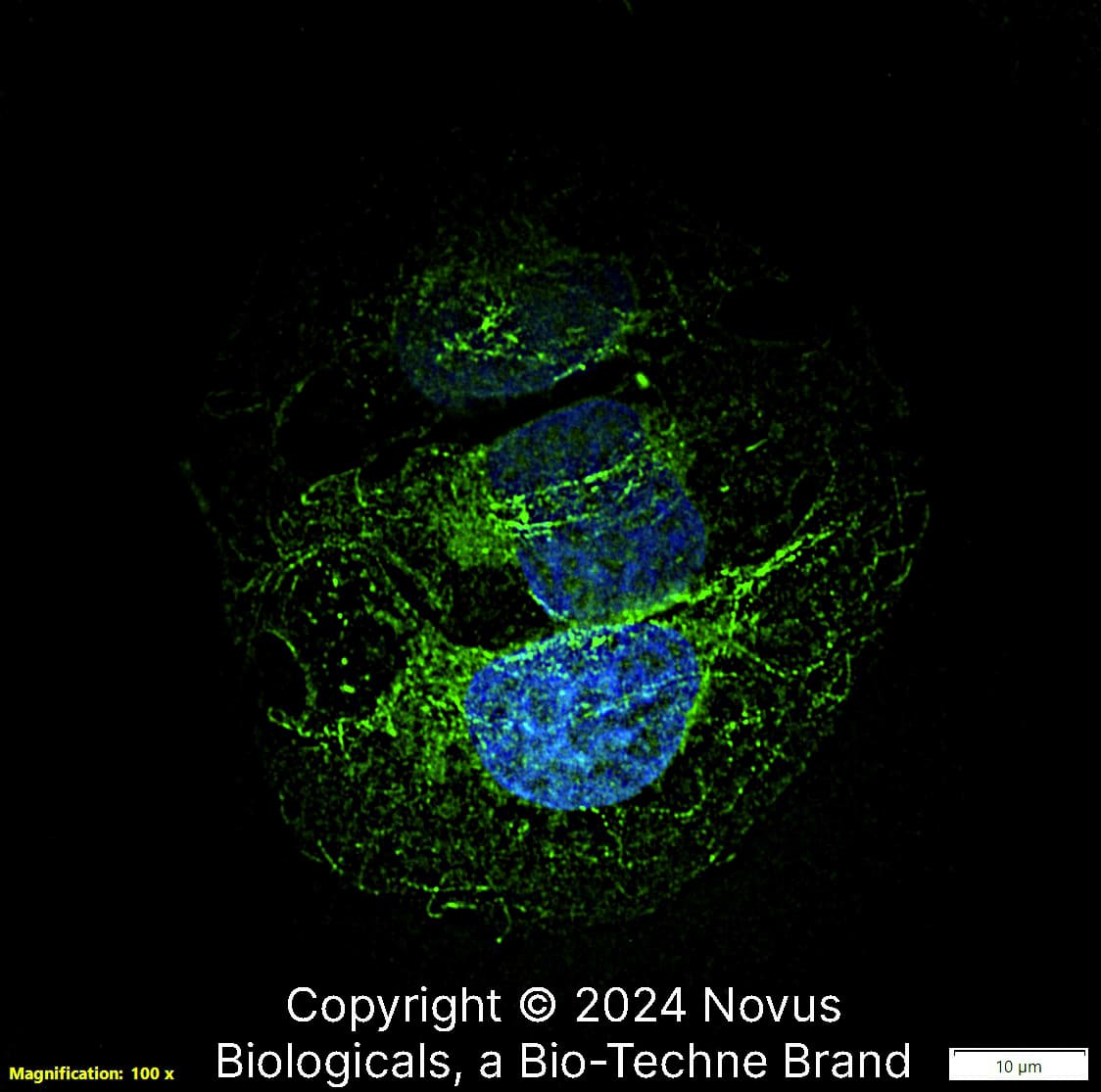
Beta Tubulin in U-2 OS Human Cell Line.
Beta Tubulin was detected in immersion fixed U-2 OS human osteosarcoma cell line using Rabbit anti-beta Tubulin Affinity Purified Polyclonal Antibody conjugated to DyLight 488 (Catalog # NB600-936G) (green) at 5 µg/mL overnight at 4C. Cells were counterstained with DAPI (blue). Cells were imaged using a 100X objective and digitally deconvolved.
Western Blot: beta Tubulin Antibody - BSA Free [NB600-936] -
Western Blot: beta Tubulin Antibody - BSA Free [NB600-936] - Dual pathway inhibition regulates TK1 protein levels & results in greater p27 protein levels than single agents alone.Single agent PLX4032 resulted in activation of p-AKT Ser473 following 24 hours of exposure at two concentrations (100 nM, 1 µM). The addition of the dual PI3K/mTOR inhibitor BEZ235 blocks p-AKT Ser473 activation & resulted in a greater increase in p27 protein levels & diminished TK1 protein levels. Image collected & cropped by CiteAb from the following publication (https://pubmed.ncbi.nlm.nih.gov/25247710), licensed under a CC-BY license. Not internally tested by Novus Biologicals.
Western Blot: beta Tubulin Antibody - BSA Free [NB600-936] -
Western Blot: beta Tubulin Antibody - BSA Free [NB600-936] - Expression of pluripotency related markers & a sphere culture of BEAS-2B under normoxia.(a) HIF-2 alpha expression was detected in the nucleus of BEAS-2B under normoxia by western blotting. Beta tubulin & TATA binding protein (TBP) were used as protein markers for the cytosol & nucleus fraction, respectively. (b) Western blotting for Oct-4. (c) For knockdown of HIF-2 alpha, BEAS-2B cells were transfected with shRNA targeting HIF-2 alpha & sh-Luc was used as control. (d) Western blotting of Nanog & a culture of BEAS-2B containing floating spheres. Representative result from two or three experiments was presented. Image collected & cropped by CiteAb from the following publication (https://pubmed.ncbi.nlm.nih.gov/27373565), licensed under a CC-BY license. Not internally tested by Novus Biologicals.
Western Blot: beta Tubulin Antibody - BSA Free [NB600-936] -
Western Blot: beta Tubulin Antibody - BSA Free [NB600-936] - Expression of AAV transgene before & after experimental autoimmune uveoretinitis (EAU) induction. (A) B10.RIII mice were evaluated by funduscopy one month after an intravitreal injection of AAV delivering sGFP-TatM013v5 or secreted GFP (sGFP). As a control we evaluated mice that received an intravitreal injection of saline. Diffuse fluorescence indicates the secretion of sGFP & sGFP-TatM013v5 in the retina. (B) Western blot from retinas harvested 14 days after IRBP immunization. Membrane was probed with anti-GFP & anti-Tubulin antibodies. Image shows expression of sGFP on both sGFP & sGFP-TatM013v5 retina lysates. (n = 2–3 retina samples from different mice per group). Image collected & cropped by CiteAb from the following publication (https://pubmed.ncbi.nlm.nih.gov/31795515), licensed under a CC-BY license. Not internally tested by Novus Biologicals.
Western Blot: beta Tubulin Antibody - BSA Free [NB600-936] -
Western Blot: beta Tubulin Antibody - BSA Free [NB600-936] - Combined V600EBRAF & mTOR inhibition results in transcriptional control of TK1 protein levels in COLO 205 cells.(A) Western blot of COLO 205 cells treated with PP242 (250 nM) & increasing PLX4032. Similar to single agent PLX4032, p-MEK, but not p-ERK, was inhibited in a PLX4032-dependent manner. Consistent with mTORC1/mTORC2 inhibition, p-AKT Ser473, but not p-AKT Thr308, was inhibited. Unlike single agent PLX4032, which resulted in concentration-dependent activation of p-rpS6, combined treatment maintained p-rpS6 levels at essentially baseline levels except at the highest PLX4032 concentration. Similarly, DUSP6 levels were inversely related to p-ERK protein levels. With combined mTOR & V600EBRAF blockade, p27 & TK1 protein levels were inversely correlated & dramatically affected by PLX4032 exposure. (B) Similarly, TK1 mRNA was significantly reduced at PLX4032 concentrations as low as 10 nM. (C) Despite elevated p27 protein levels, p27 mRNA was unaffected by combined mTOR-V600EBRAF inhibition. Image collected & cropped by CiteAb from the following publication (https://pubmed.ncbi.nlm.nih.gov/25247710), licensed under a CC-BY license. Not internally tested by Novus Biologicals.
Western Blot: beta Tubulin Antibody - BSA Free [NB600-936] -
Western Blot: beta Tubulin Antibody - BSA Free [NB600-936] - [18F]-FLT PET reflects BEZ235-dependent inhibition of PI3K/mTOR activity in PLX4720 treated COLO 205 xenografts. Xenograft-bearing mice were imaged with [18F]-FLT PET on treatment day 4. (A) [18F]-FLT uptake was diminished in the combination treatment cohort relative to vehicle (p = 0.0087), but not single agent PLX4720- or BEZ235-treated cohorts. (B) Western blot of xenograft tissue harvested immediately following imaging illustrated elevated p-ERK & p-rpS6 levels in PLX4720-treated mice. Combining PLX4032 with BEZ235 resulted in reduced p-ERK & p-rpS6 protein levels. (C) TK1 levels, as measured by IHC, were reduced only in the combination treatment group in agreement with [18F]-FLT PET. (D) Consistent with in vitro studies, diminished TK1 levels, & consequently [18F]-FLT PET, correlated with elevated p27 that was elevated only in the combination treated group. Image collected & cropped by CiteAb from the following publication (https://pubmed.ncbi.nlm.nih.gov/25247710), licensed under a CC-BY license. Not internally tested by Novus Biologicals.
Western Blot: beta Tubulin Antibody - BSA Free [NB600-936] -
Western Blot: beta Tubulin Antibody - BSA Free [NB600-936] - PLX4720 exposure does not affect [18F]-FLT PET in COLO 205 xenografts, despite evidence of target inhibition & diminished [18F]-FDG uptake.(A) Representative transverse [18F]-FLT & [18F]-FDG PET images acquired after three daily treatments with vehicle or 60 mg/kg PLX4720 (tumor indicated by arrowhead). (B) Quantification of PET data illustrated similar [18F]-FLT uptake in vehicle-treated & PLX4720-treated tumors. Unlike [18F]-FLT PET, PLX4720 exposure elicited a significant reduction in [18F]-FDG uptake (p = 0.0006). (C) Western blot analysis of vehicle- & PLX4720-treated tumor tissue confirmed that PLX4720 had no effect on TK1 protein levels in agreement with [18F]-FLT PET. Target inhibition as measured by p-MEK levels was observed. However, similar to in vitro studies, PLX4720-treated COLO 205 xenografts exhibited elevated p-ERK & p-rpS6 protein levels relative to vehicle controls. Image collected & cropped by CiteAb from the following publication (https://pubmed.ncbi.nlm.nih.gov/25247710), licensed under a CC-BY license. Not internally tested by Novus Biologicals.
Western Blot: beta Tubulin Antibody - BSA Free [NB600-936] -
Western Blot: beta Tubulin Antibody - BSA Free [NB600-936] - Deletion of Phb1 affects lipid metabolism.Western blot (a) & quantification (b) of Acetyl-CoA carboxylase (ACC) & phosphorylated ACC (p-ACC) expression at P20. N = 6–7 animals per genotype. Unpaired two-tailed t-test [p-ACC (t = 0.4627, df = 11, p = 0.021), ACC (t = 1.355, df = 11, p = 0.26)]. Western blot (c) & quantification (d) of ACC & p-ACC expression at P40. N = 6–8 animals per genotype [p-ACC (t = 0.5447, df = 12, p = 0.42), ACC (t = 1.153, df = 12, p = 0.17)]. Unpaired two-tailed t-test. By RT-qPCR, we identified a significant downregulation of many enzymes involved with lipid biosynthesis at both P20 (e) & P40 (f): sterol regulatory element-binding protein 1 (Srebp1), 3-hydroxy-3-methylglutaryl-CoA reductase (Hmgcr), ATP citrate lyase (Acly), fatty acid synthase (FASN), acetyl-CoA carboxylase 2 (ACC2), N = 5 animals per genotype. Unpaired two-tailed t-test P20 [Srebp1 (t = 3.26, df = 8, p = 0.012), Hmgcr (t = 7.63, df = 8, p = 0.000061), Acly (t = 4.418, df = 8, 0.0022), FASN (t = 4.109, df = 8, p = 0.0034), ACC2 (t = 3.408, df = 8, p = 0.0092)]; P40 [Srebp1 (t = 7.551, df = 8, p = 0.000066), Hmgcr (t = 5.091, df = 8, p = 0.00094), Acly (t = 4.934, df = 8, p = 0.0011), FASN (t = 7.186, df = 8, p = 0.000094), ACC2 (t = 2.697, df = 8, p = 0.027)]. Data are presented as mean ± SEM. *p < 0.05; **p < 0.01; ***p < 0.001. n.s. non-significant. Image collected & cropped by CiteAb from the following publication (https://pubmed.ncbi.nlm.nih.gov/34078899), licensed under a CC-BY license. Not internally tested by Novus Biologicals.
Western Blot: beta Tubulin Antibody - BSA Free [NB600-936] -
Western Blot: beta Tubulin Antibody - BSA Free [NB600-936] - TK1 protein levels do not reflect p-ERK attenuation following inhibition of V600EBRAF inhibition in COLO 205 cells.COLO 205 cells were collected 48 hours of PLX 4032 exposure at 10 nM, 100 nM, 500 nM, 1 µM, or 5 µM. (A) Western blot analysis demonstrated target inhibition of p-MEK despite increased p-ERK levels. PI3K-mTOR signaling was elevated in a PLX 4032-dependent manner as exhibited by a steady rise in p-rpS6 levels. The ERK-phosphatase DUSP6 decreased in conjunction with mTOR signaling & was inversely proportional to p-ERK levels. A slight increase in p27 levels were observed concomitantly with only modest changes in TK1 levels, except at the highest dose of PLX4032. (B) Decreased TK1 mRNA levels were observed at all drug concentrations above 10 nM (p<0.05). Image collected & cropped by CiteAb from the following publication (https://pubmed.ncbi.nlm.nih.gov/25247710), licensed under a CC-BY license. Not internally tested by Novus Biologicals.

![Immunocytochemistry/ Immunofluorescence: beta Tubulin Antibody - BSA Free [NB600-936] Immunocytochemistry/ Immunofluorescence: beta Tubulin Antibody - BSA Free [NB600-936]](https://resources.bio-techne.com/images/products/beta-Tubulin-Antibody-Immunocytochemistry-Immunofluorescence-NB600-936-img0021.jpg)
![Simple Western: beta Tubulin AntibodyBSA Free [NB600-936] Simple Western: beta Tubulin AntibodyBSA Free [NB600-936]](https://resources.bio-techne.com/images/products/beta-Tubulin-Antibody-Simple-Western-NB600-936-img0016.jpg)
![Western Blot: beta Tubulin AntibodyBSA Free [NB600-936] Western Blot: beta Tubulin AntibodyBSA Free [NB600-936]](https://resources.bio-techne.com/images/products/beta-Tubulin-Antibody-Western-Blot-NB600-936-img0006.jpg)
![Western Blot: beta Tubulin AntibodyBSA Free [NB600-936] Western Blot: beta Tubulin AntibodyBSA Free [NB600-936]](https://resources.bio-techne.com/images/products/beta-Tubulin-Antibody-Western-Blot-NB600-936-img0020.jpg)
![Immunocytochemistry/ Immunofluorescence: beta Tubulin Antibody - BSA Free [NB600-936] Immunocytochemistry/ Immunofluorescence: beta Tubulin Antibody - BSA Free [NB600-936]](https://resources.bio-techne.com/images/products/beta-Tubulin-Antibody-Immunocytochemistry-Immunofluorescence-NB600-936-img0023.jpg)
![Immunohistochemistry-Paraffin: beta Tubulin Antibody - BSA Free [NB600-936] Immunohistochemistry-Paraffin: beta Tubulin Antibody - BSA Free [NB600-936]](https://resources.bio-techne.com/images/products/beta-Tubulin-Antibody-Immunohistochemistry-Paraffin-NB600-936-img0011.jpg)
![Western Blot: beta Tubulin AntibodyBSA Free [NB600-936] Western Blot: beta Tubulin AntibodyBSA Free [NB600-936]](https://resources.bio-techne.com/images/products/beta-Tubulin-Antibody-Western-Blot-NB600-936-img0004.jpg)
![Western Blot: beta Tubulin AntibodyBSA Free [NB600-936] Western Blot: beta Tubulin AntibodyBSA Free [NB600-936]](https://resources.bio-techne.com/images/products/beta-Tubulin-Antibody-Western-Blot-NB600-936-img0024.jpg)
![Immunocytochemistry/ Immunofluorescence: beta Tubulin Antibody - BSA Free [NB600-936] Immunocytochemistry/ Immunofluorescence: beta Tubulin Antibody - BSA Free [NB600-936]](https://resources.bio-techne.com/images/products/beta-Tubulin-Antibody-Immunocytochemistry-Immunofluorescence-NB600-936-img0015.jpg)
![Immunocytochemistry/ Immunofluorescence: beta Tubulin Antibody - BSA Free [NB600-936] Immunocytochemistry/ Immunofluorescence: beta Tubulin Antibody - BSA Free [NB600-936]](https://resources.bio-techne.com/images/products/beta-Tubulin-Antibody-Immunocytochemistry-Immunofluorescence-NB600-936-img0001.jpg)
![Immunocytochemistry/ Immunofluorescence: beta Tubulin Antibody - BSA Free [NB600-936] Immunocytochemistry/ Immunofluorescence: beta Tubulin Antibody - BSA Free [NB600-936]](https://resources.bio-techne.com/images/products/beta-Tubulin-Antibody-Immunocytochemistry-Immunofluorescence-NB600-936-img0022.jpg)
![Immunohistochemistry-Paraffin: beta Tubulin Antibody - BSA Free [NB600-936] Immunohistochemistry-Paraffin: beta Tubulin Antibody - BSA Free [NB600-936]](https://resources.bio-techne.com/images/products/beta-Tubulin-Antibody-Immunohistochemistry-Paraffin-NB600-936-img0014.jpg)
![Immunohistochemistry-Paraffin: beta Tubulin Antibody - BSA Free [NB600-936] Immunohistochemistry-Paraffin: beta Tubulin Antibody - BSA Free [NB600-936]](https://resources.bio-techne.com/images/products/beta-Tubulin-Antibody-Immunohistochemistry-Paraffin-NB600-936-img0013.jpg)
![Immunohistochemistry-Paraffin: beta Tubulin Antibody - BSA Free [NB600-936] Immunohistochemistry-Paraffin: beta Tubulin Antibody - BSA Free [NB600-936]](https://resources.bio-techne.com/images/products/beta-Tubulin-Antibody-Immunohistochemistry-Paraffin-NB600-936-img0012.jpg)


![Western Blot: beta Tubulin Antibody - BSA Free [NB600-936] - beta Tubulin Antibody - BSA Free](https://resources.bio-techne.com/images/products/nb600-936_rabbit-polyclonal-beta-tubulin-antibody-310202415175297.jpg)
![Western Blot: beta Tubulin Antibody - BSA Free [NB600-936] - beta Tubulin Antibody - BSA Free](https://resources.bio-techne.com/images/products/nb600-936_rabbit-polyclonal-beta-tubulin-antibody-310202415334929.jpg)
![Western Blot: beta Tubulin Antibody - BSA Free [NB600-936] - beta Tubulin Antibody - BSA Free](https://resources.bio-techne.com/images/products/nb600-936_rabbit-polyclonal-beta-tubulin-antibody-310202415384143.jpg)
![Western Blot: beta Tubulin Antibody - BSA Free [NB600-936] - beta Tubulin Antibody - BSA Free](https://resources.bio-techne.com/images/products/nb600-936_rabbit-polyclonal-beta-tubulin-antibody-310202416171469.jpg)
![Western Blot: beta Tubulin Antibody - BSA Free [NB600-936] - beta Tubulin Antibody - BSA Free](https://resources.bio-techne.com/images/products/nb600-936_rabbit-polyclonal-beta-tubulin-antibody-31020241617144.jpg)
![Western Blot: beta Tubulin Antibody - BSA Free [NB600-936] - beta Tubulin Antibody - BSA Free](https://resources.bio-techne.com/images/products/nb600-936_rabbit-polyclonal-beta-tubulin-antibody-310202416171490.jpg)
![Western Blot: beta Tubulin Antibody - BSA Free [NB600-936] - beta Tubulin Antibody - BSA Free](https://resources.bio-techne.com/images/products/nb600-936_rabbit-polyclonal-beta-tubulin-antibody-31020241616030.jpg)
![Western Blot: beta Tubulin Antibody - BSA Free [NB600-936] - beta Tubulin Antibody - BSA Free](https://resources.bio-techne.com/images/products/nb600-936_rabbit-polyclonal-beta-tubulin-antibody-31020241616031.jpg)
![Western Blot: beta Tubulin Antibody - BSA Free [NB600-936] - beta Tubulin Antibody - BSA Free](https://resources.bio-techne.com/images/products/nb600-936_rabbit-polyclonal-beta-tubulin-antibody-310202416175297.jpg)