Calreticulin Antibody - BSA Free
Novus Biologicals, part of Bio-Techne | Catalog # NB600-101

![Western Blot: Calreticulin Antibody - BSA Free [NB600-101] Knockdown Validated: Calreticulin Antibody - BSA Free [NB600-101]](https://resources.bio-techne.com/images/products/Calreticulin-Antibody-Knockdown-Validated-NB600-101-img0011.jpg)
Conjugate
Catalog #
Key Product Details
Validated by
Knockout/Knockdown
Species Reactivity
Validated:
Human, Mouse, Rat, Bovine, Hamster, Primate
Cited:
Human, Mouse, Rat, Bovine, Hamster, Primate
Applications
Validated:
Block/Neutralize, Dot Blot, Electron Microscopy, Flow Cytometry, Immunocytochemistry/ Immunofluorescence, Immunohistochemistry, Immunohistochemistry-Paraffin, Immunoprecipitation, Knockdown Validated, Protein Array, Simple Western, Western Blot
Cited:
Block/Neutralize, Cytometric Bead Assay Standard, Electron Microscopy, Flow Cytometry, IF/IHC, Immunocytochemistry/ Immunofluorescence, Immunoprecipitation, Protein Array, Western Blot
Label
Unconjugated
Antibody Source
Polyclonal Rabbit IgG
Format
BSA Free
Concentration
1.0 mg/ml
Product Specifications
Immunogen
Calreticulin Antibody was developed against a fusion protein to mouse Calreticulin
Reactivity Notes
Other species not tested.
Localization
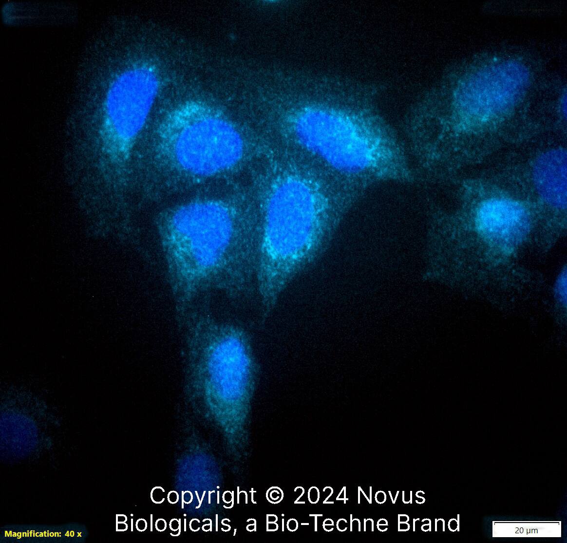
Calreticulin staining is localized to the ER in most cell types and under most conditions.
Marker
Endoplasmic Reticulum Marker
Clonality
Polyclonal
Host
Rabbit
Isotype
IgG
Theoretical MW
48 kDa.
Disclaimer note: The observed molecular weight of the protein may vary from the listed predicted molecular weight due to post translational modifications, post translation cleavages, relative charges, and other experimental factors.
Disclaimer note: The observed molecular weight of the protein may vary from the listed predicted molecular weight due to post translational modifications, post translation cleavages, relative charges, and other experimental factors.
Scientific Data Images for Calreticulin Antibody - BSA Free
Western Blot: Calreticulin Antibody - BSA Free [NB600-101]
Western Blot: Calreticulin Antibody [NB600-101] - Western blot shows lysates of HeLa human cervical epithelial carcinoma parental cell line and Calreticulin knockout (KO) HeLa cell line. PVDF membrane was probed with 1:1500 of Rabbit Anti-Human Calreticulin Polyclonal Antibody (Catalog # NB600-101) followed by HRP-conjugated Anti-Rabbit IgG Secondary Antibody (Catalog #HAF008). Specific band was detected for Calreticulin at approximately 55 kDa (as indicated) in the parental HeLa cell line, but is not detectable in the knockout HeLa cell line. This experiment was conducted under reducing conditions.Western Blot: Calreticulin AntibodyBSA Free [NB600-101]
Western Blot: Calreticulin Antibody [NB600-101] - Human kidney lysate.Immunocytochemistry/ Immunofluorescence: Calreticulin Antibody - BSA Free [NB600-101]
Immunocytochemistry/Immunofluorescence: Calreticulin Antibody [NB600-101] - Immunofluorescence staining of Calreticulin in HCT15 colon cancer cells using NB600-101. Secondary antibody was conjugated with Alexa Fluor 488. Photo courtesy of Dr. Birkenkamp Demtroeder, Arhus University Hospital.Applications for Calreticulin Antibody - BSA Free
Application
Recommended Usage
Flow Cytometry
reported in scientific literature (PMID 19171874)
Immunocytochemistry/ Immunofluorescence
1:50-1:250
Immunohistochemistry
1:50 - 1:200
Immunohistochemistry-Paraffin
1:50 -1:200
Immunoprecipitation
reported in scientific literature (PMID 15161937)
Knockdown Validated
1:1500
Protein Array
reported in scientific literature (PMID 17897946)
Simple Western
1:50
Western Blot
1:1000
Application Notes
In Western blot, a band is observed at ~55 kDa. The observed molecular weight of the protein may vary from the listed predicted molecular weight due to post translational modifications, post translation cleavages, relative charges, and other experimental factors. In Simple Western only 10 - 15 uL of the recommended dilution is used per data point.
See Simple Western Antibody Database for Simple Western validation: Tested in HeLa lysate 0.5 mg/mL, separated by Size, antibody dilution of 1:50, apparent MW was 64 kDa. Separated by Size-Wes, Sally Sue/Peggy Sue.
See Simple Western Antibody Database for Simple Western validation: Tested in HeLa lysate 0.5 mg/mL, separated by Size, antibody dilution of 1:50, apparent MW was 64 kDa. Separated by Size-Wes, Sally Sue/Peggy Sue.
Formulation, Preparation, and Storage
Purification
Immunogen affinity purified
Formulation
PBS
Format
BSA Free
Preservative
0.02% Sodium Azide
Concentration
1.0 mg/ml
Shipping
The product is shipped with polar packs. Upon receipt, store it immediately at the temperature recommended below.
Stability & Storage
Aliquot and store at -20C or -80C. Avoid freeze-thaw cycles.
Background: Calreticulin
Given its role in multiple biological processes, it makes sense that calreticulin is implicated in both healthy and disease states. Studies have found that calreticulin mutations were present in patients with myeloproliferative neoplasms (MFN) and essential thrombocythaemia (ET) (4). Calreticulin expression is typically upregulated in most cancer lines, however it is downregulated in some tissues including cervical carcinomas, prostate cancer, and human colon adenocarcinoma (3). High expression of calreticulin on cancer cells is related to tumor cell phagocytosis and is often correlated with, and counteracted by, elevated CD47 expression to prevent cancer cell phagocytosis (3). Calreticulin mutations can serve as a major diagnostic biomarker for MFN and ET and additionally the calreticulin gene may be a potential target for cancer therapeutics (3,4).
References
1. Michalak, M., Groenendyk, J., Szabo, E., Gold, L. I., & Opas, M. (2009). Calreticulin, a multi-process calcium-buffering chaperone of the endoplasmic reticulum. The Biochemical Journal. https://doi.org/10.1042/BJ20081847
2. Fucikova, J., Spisek, R., Kroemer, G., & Galluzzi, L. (2021). Calreticulin and cancer. Cell Research. https://doi.org/10.1038/s41422-020-0383-9
3. Sun, J., Mu, H., Dai, K., & Yi, L. (2017). Calreticulin: a potential anti-cancer therapeutic target. Die Pharmazie. https://doi.org/10.1691/ph.2017.7031
4. Prins, D., Gonzalez Arias, C., Klampfl, T., Grinfeld, J., & Green, A. R. (2020). Mutant Calreticulin in the Myeloproliferative Neoplasms. HemaSphere. https://doi.org/10.1097/HS9.0000000000000333
Alternate Names
Autoantigen Ro, CALR, Calregulin, cC1qR, CRT, SSA, Vasostatin
Gene Symbol
CALR
UniProt
Additional Calreticulin Products
Product Documents for Calreticulin Antibody - BSA Free
Product Specific Notices for Calreticulin Antibody - BSA Free
This product is for research use only and is not approved for use in humans or in clinical diagnosis. Primary Antibodies are guaranteed for 1 year from date of receipt.
Loading...
Loading...
Loading...
Loading...
Loading...
Loading...
![Western Blot: Calreticulin AntibodyBSA Free [NB600-101] Western Blot: Calreticulin AntibodyBSA Free [NB600-101]](https://resources.bio-techne.com/images/products/Calreticulin-Antibody-Western-Blot-NB600-101-img0006.jpg)
![Immunocytochemistry/ Immunofluorescence: Calreticulin Antibody - BSA Free [NB600-101] Immunocytochemistry/ Immunofluorescence: Calreticulin Antibody - BSA Free [NB600-101]](https://resources.bio-techne.com/images/products/Calreticulin-Antibody-Immunocytochemistry-Immunofluorescence-NB600-101-img0005.jpg)
![Immunohistochemistry-Paraffin: Calreticulin Antibody - BSA Free [NB600-101] Immunohistochemistry-Paraffin: Calreticulin Antibody - BSA Free [NB600-101]](https://resources.bio-techne.com/images/products/Calreticulin-Antibody-Immunohistochemistry-Paraffin-NB600-101-img0010.jpg)
![Immunocytochemistry/ Immunofluorescence: Calreticulin Antibody - BSA Free [NB600-101] Immunocytochemistry/ Immunofluorescence: Calreticulin Antibody - BSA Free [NB600-101]](https://resources.bio-techne.com/images/products/Calreticulin-Antibody-Immunocytochemistry-Immunofluorescence-NB600-101-img0007.jpg)
![Immunohistochemistry-Paraffin: Calreticulin Antibody - BSA Free [NB600-101] Immunohistochemistry-Paraffin: Calreticulin Antibody - BSA Free [NB600-101]](https://resources.bio-techne.com/images/products/Calreticulin-Antibody-Immunohistochemistry-Paraffin-NB600-101-img0009.jpg)
![Simple Western: Calreticulin AntibodyBSA Free [NB600-101] Simple Western: Calreticulin AntibodyBSA Free [NB600-101]](https://resources.bio-techne.com/images/products/Calreticulin-Antibody-Simple-Western-NB600-101-img0008.jpg)
