E-Cadherin Antibody (7H12)
Novus Biologicals, part of Bio-Techne | Catalog # NBP2-19051

Key Product Details
Validated by
Knockout/Knockdown, Biological Validation
Species Reactivity
Validated:
Human, Mouse, Monkey
Cited:
Human, Mouse
Applications
Validated:
ELISA, Flow Cytometry, Immunocytochemistry/ Immunofluorescence, Immunohistochemistry, Immunohistochemistry-Paraffin, Multiplex Immunofluorescence, Western Blot
Cited:
IF/IHC, Immunocytochemistry/ Immunofluorescence, Immunohistochemistry-Frozen, Immunohistochemistry-Paraffin, Western Blot
Label
Unconjugated
Antibody Source
Monoclonal Mouse IgG1 Clone # 7H12
Concentration
This product is unpurified. The exact concentration of antibody is not quantifiable.
Product Specifications
Immunogen
Purified recombinant fragment of human E-Cadherin expressed in E. Coli.
Reactivity Notes
Please note that this antibody is reactive to Mouse and derived from the same host, Mouse. Mouse-On-Mouse blocking reagent may be needed for IHC and ICC experiments to reduce high background signal. You can find these reagents under catalog numbers PK-2200-NB and MP-2400-NB. Please contact Technical Support if you have any questions.
Marker
Epithelial Cell Marker, Adherens Junctions Marker
Clonality
Monoclonal
Host
Mouse
Isotype
IgG1
Theoretical MW
135 kDa.
Disclaimer note: The observed molecular weight of the protein may vary from the listed predicted molecular weight due to post translational modifications, post translation cleavages, relative charges, and other experimental factors.
Disclaimer note: The observed molecular weight of the protein may vary from the listed predicted molecular weight due to post translational modifications, post translation cleavages, relative charges, and other experimental factors.
Scientific Data Images for E-Cadherin Antibody (7H12)
Detection of E-Cadherin in Human Breast via Multiplex Immunofluorescence staining on COMET™
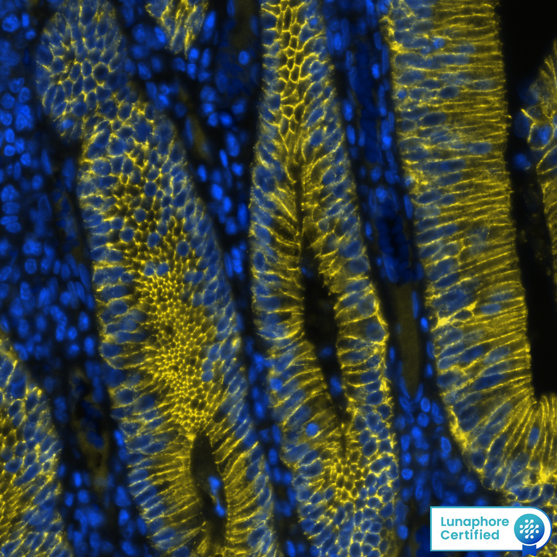
E-Cadherin was detected in immersion fixed paraffin-embedded sections of human breast using Mouse Anti-Human E-Cadherin Monoclonal Antibody (Novus Catalog # NBP2-19051) at 1:1000 at 37 ° Celsius for 4 minutes. Before incubation with the primary antibody, tissue underwent an all-in-one dewaxing and antigen retrieval preprocessing using PreTreatment Module (PT Module) and Dewax and HIER Buffer H (pH 9). Tissue was stained using the Alexa Fluor™ Plus 647 Goat anti-Mouse IgG Secondary Antibody at 1:200 at 37 ° Celsius for 2 minutes. (Yellow; Lunaphore Catalog # DR647MS) and counterstained with DAPI (blue; Lunaphore Catalog # DR100). Specific staining was localized to the membrane. Protocol available in COMET™ Panel Builder.Detection of E-Cadherin in Human Liver via Multiplex Immunofluorescence staining on COMET™
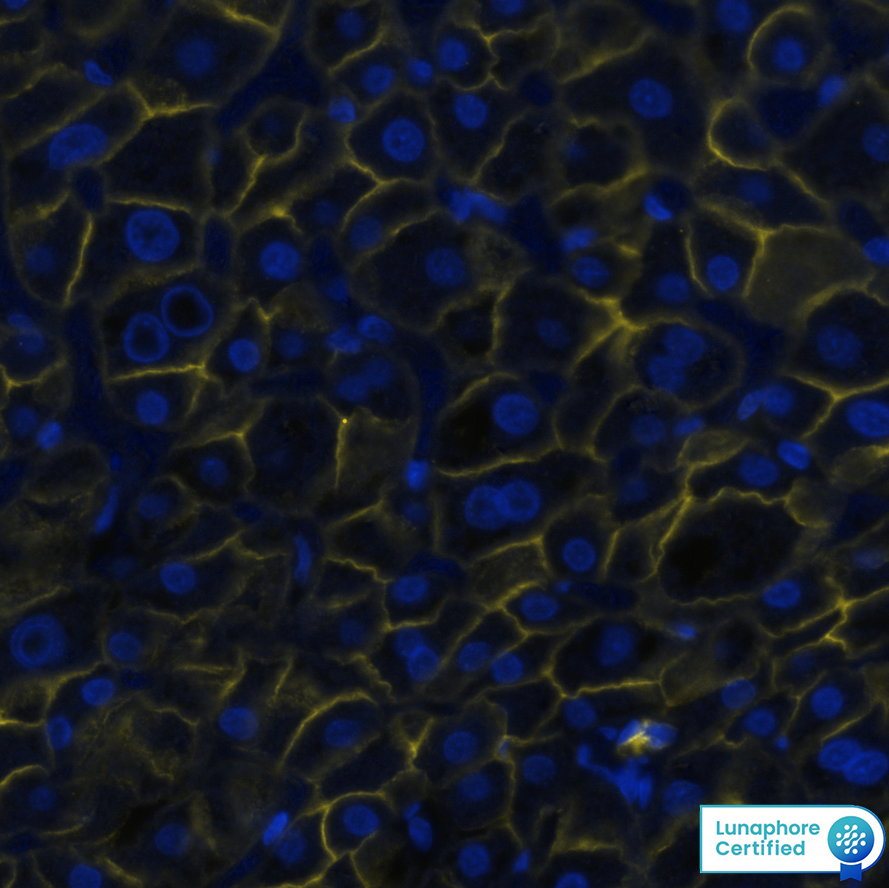
E-Cadherin was detected in immersion fixed paraffin-embedded sections of human liver using Mouse Anti-Human E-Cadherin Monoclonal Antibody (Catalog # NBP2-19051) at 1:1000 at 37 ° Celsius for 4 minutes. Before incubation with the primary antibody, tissue underwent an all-in-one dewaxing and antigen retrieval preprocessing using PreTreatment Module (PT Module) and Dewax and HIER Buffer H (pH 9). Tissue was stained using the Alexa Fluor™ 647 Goat anti-Mouse IgG Secondary Antibody at 1:200 at 37 ° Celsius for 2 minutes. (Yellow; Lunaphore Catalog # DR647MS) and counterstained with DAPI (blue; Lunaphore Catalog # DR100). Specific staining was localized to the membrane. Protocol available in COMET™ Panel Builder.Detection of E-Cadherin in Human Colon Cancer via Multiplex Immunofluorescence staining on COMET™
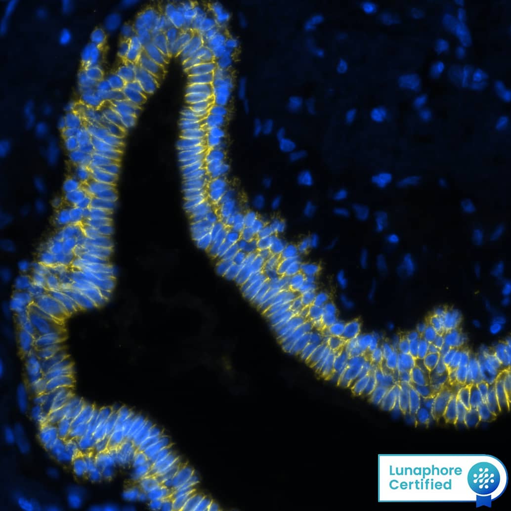
E-Cadherin was detected in immersion fixed paraffin-embedded sections of human colon cancer using Mouse Anti-Human E-Cadherin Monoclonal Antibody (Catalog # NBP2-19051) at 1:1000 at 37 ° Celsius for 4 minutes. Before incubation with the primary antibody, tissue underwent an all-in-one dewaxing and antigen retrieval preprocessing using PreTreatment Module (PT Module) and Dewax and HIER Buffer H (pH 9). Tissue was stained using the Alexa Fluor™ 647 Goat anti-Mouse IgG Secondary Antibody at 1:200 at 37 ° Celsius for 2 minutes. (Yellow; Lunaphore Catalog # DR647MS) and counterstained with DAPI (blue; Lunaphore Catalog # DR100). Specific staining was localized to the membrane. Protocol available in COMET™ Panel Builder.Applications for E-Cadherin Antibody (7H12)
Application
Recommended Usage
ELISA
1:10000
Flow Cytometry
1:200-1:400
Immunocytochemistry/ Immunofluorescence
1:10-1:500
Immunohistochemistry
1:200-1:1000
Immunohistochemistry-Paraffin
1:200-1:1000
Multiplex Immunofluorescence
1:1000
Western Blot
1:500-1:2000
Application Notes
Use in ICC/IF reported in scientific literature (PMID: 20605541).
Reviewed Applications
Read 7 reviews rated 4.4 using NBP2-19051 in the following applications:
Formulation, Preparation, and Storage
Purification
Unpurified
Formulation
Ascites
Preservative
0.03% Sodium Azide
Concentration
This product is unpurified. The exact concentration of antibody is not quantifiable.
Shipping
The product is shipped with polar packs. Upon receipt, store it immediately at the temperature recommended below.
Stability & Storage
Store at 4C short term. Aliquot and store at -20C long term. Avoid freeze-thaw cycles.
Background: E-Cadherin
Alternate Names
Arc-1, CAD1, Cadherin-1, CD324, CDH1, Cell-CAM120/80, ECAD, ECadherin, L-CAM, Uvomorulin
Entrez Gene IDs
999 (Human)
Gene Symbol
CDH1
UniProt
Additional E-Cadherin Products
Product Documents for E-Cadherin Antibody (7H12)
Product Specific Notices for E-Cadherin Antibody (7H12)
This product is for research use only and is not approved for use in humans or in clinical diagnosis. Primary Antibodies are guaranteed for 1 year from date of receipt.
Loading...
Loading...
Loading...
Loading...
Loading...


![Immunohistochemistry-Paraffin: E-Cadherin Antibody (7H12) - BSA Free [NBP2-19051] Immunohistochemistry-Paraffin: E-Cadherin Antibody (7H12) - BSA Free [NBP2-19051]](https://resources.bio-techne.com/images/products/E-Cadherin-Antibody-7H12-Immunohistochemistry-Paraffin-NBP2-19051-img0011.jpg)

![Western Blot: E-Cadherin Antibody (7H12)BSA Free [NBP2-19051] Western Blot: E-Cadherin Antibody (7H12)BSA Free [NBP2-19051]](https://resources.bio-techne.com/images/products/E-Cadherin-Antibody-7H12-Western-Blot-NBP2-19051-img0013.jpg)
![Western Blot: E-Cadherin Antibody (7H12)BSA Free [NBP2-19051] Western Blot: E-Cadherin Antibody (7H12)BSA Free [NBP2-19051]](https://resources.bio-techne.com/images/products/E-Cadherin-Antibody-7H12-Western-Blot-NBP2-19051-img0002.jpg)
![Immunocytochemistry/ Immunofluorescence: E-Cadherin Antibody (7H12) - BSA Free [NBP2-19051] Immunocytochemistry/ Immunofluorescence: E-Cadherin Antibody (7H12) - BSA Free [NBP2-19051]](https://resources.bio-techne.com/images/products/E-Cadherin-Antibody-7H12-Immunocytochemistry-Immunofluorescence-NBP2-19051-img0010.jpg)
![Western Blot: E-Cadherin Antibody (7H12)BSA Free [NBP2-19051] Western Blot: E-Cadherin Antibody (7H12)BSA Free [NBP2-19051]](https://resources.bio-techne.com/images/products/E-Cadherin-Antibody-7H12-Western-Blot-NBP2-19051-img0014.jpg)
![Immunocytochemistry/ Immunofluorescence: E-Cadherin Antibody (7H12) - BSA Free [NBP2-19051] Immunocytochemistry/ Immunofluorescence: E-Cadherin Antibody (7H12) - BSA Free [NBP2-19051]](https://resources.bio-techne.com/images/products/E-Cadherin-Antibody-7H12-Immunocytochemistry-Immunofluorescence-NBP2-19051-img0009.jpg)
![Immunohistochemistry-Paraffin: E-Cadherin Antibody (7H12) - BSA Free [NBP2-19051] Immunohistochemistry-Paraffin: E-Cadherin Antibody (7H12) - BSA Free [NBP2-19051]](https://resources.bio-techne.com/images/products/E-Cadherin-Antibody-7H12-Immunohistochemistry-Paraffin-NBP2-19051-img0012.jpg)
![Flow Cytometry: E-Cadherin Antibody (7H12) - BSA Free [NBP2-19051] Flow Cytometry: E-Cadherin Antibody (7H12) - BSA Free [NBP2-19051]](https://resources.bio-techne.com/images/products/E-Cadherin-Antibody-7H12-Flow-Cytometry-NBP2-19051-img0004.jpg)
![Western Blot: E-Cadherin Antibody (7H12)BSA Free [NBP2-19051] Western Blot: E-Cadherin Antibody (7H12)BSA Free [NBP2-19051]](https://resources.bio-techne.com/images/products/E-Cadherin-Antibody-7H12-Western-Blot-NBP2-19051-img0015.jpg)
![Immunohistochemistry-Paraffin: E-Cadherin Antibody (7H12) - BSA Free [NBP2-19051] Immunohistochemistry-Paraffin: E-Cadherin Antibody (7H12) - BSA Free [NBP2-19051]](https://resources.bio-techne.com/images/products/E-Cadherin-Antibody-7H12-Immunohistochemistry-Paraffin-NBP2-19051-img0003.jpg)
![Western Blot: E-Cadherin Antibody (7H12) - BSA Free [NBP2-19051] - E-Cadherin Antibody (7H12) - BSA Free](https://resources.bio-techne.com/images/products/nbp2-19051_mouse-monoclonal-e-cadherin-antibody-7h12-210202423454897.jpg)