Human AHR Antibody
R&D Systems, part of Bio-Techne | Catalog # AF6185

Key Product Details
Validated by
Species Reactivity
Validated:
Cited:
Applications
Validated:
Cited:
Label
Antibody Source
Product Specifications
Immunogen
Asn704-Leu848
Accession # P35869
Specificity
Clonality
Host
Isotype
Scientific Data Images for Human AHR Antibody
Detection of Human AHR by Western Blot.
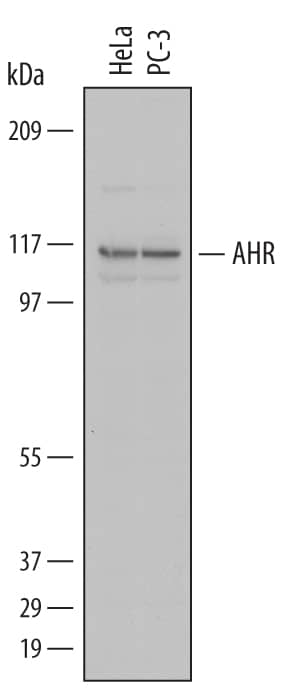
Western blot shows lysates of HeLa human cervical epithelial carcinoma cell line and PC-3 human prostate cancer cell line. PVDF Membrane was probed with 1 µg/mL of Sheep Anti-Human AHR Antigen Affinity-purified Polyclonal Antibody (Catalog # AF6185) followed by HRP-conjugated Anti-Sheep IgG Secondary Antibody (Catalog # HAF016). A specific band was detected for AHR at approximately 110 kDa (as indicated). This experiment was conducted under reducing conditions and using Immunoblot Buffer Group 1.AHR in HeLa Human Cell Line.
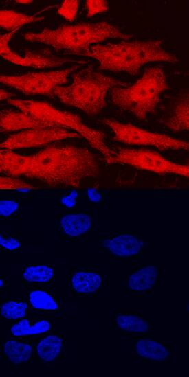
AHR was detected in immersion fixed HeLa human cervical epithelial carcinoma cell line using Sheep Anti-Human AHR Antigen Affinity-purified Polyclonal Antibody (Catalog # AF6185) at 10 µg/mL for 3 hours at room temperature. Cells were stained using the NorthernLights™ 557-conjugated Anti-Sheep IgG Secondary Antibody (red, upper panel; Catalog # NL010) and counterstained with DAPI (blue, lower panel). Specific staining was localized to nuclei and cytoplasm. View our protocol for Fluorescent ICC Staining of Cells on Coverslips.Detection of AHR in HeLa Human Cell Line by Flow Cytometry.
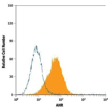
HeLa human cervical epithelial carcinoma cell line was stained with Sheep Anti-Human AHR Antigen Affinity-purified Polyclonal Antibody (Catalog # AF6185, filled histogram) or isotype control antibody (Catalog # 5-001-A, open histogram), followed by Phycoerythrin-conjugated Anti-Sheep IgG Secondary Antibody (Catalog # F0126). To facilitate intracellular staining, cells were fixed with paraformaldehyde and permeabilized with saponin.Applications for Human AHR Antibody
CyTOF-ready
Immunocytochemistry
Sample: Immersion fixed HeLa human cervical epithelial carcinoma cell line
Intracellular Staining by Flow Cytometry
Sample: HeLa human cervical epithelial carcinoma cell line fixed with paraformaldehyde and permeabilized with saponin
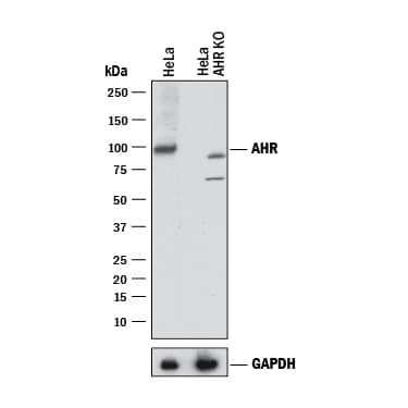
Knockout Validated
Simple Western
Sample: PC‑3 human prostate cancer cell line
Western Blot
Sample: HeLa human cervical epithelial carcinoma cell line and PC‑3 human prostate cancer cell line
Formulation, Preparation, and Storage
Purification
Reconstitution
Formulation
Shipping
Stability & Storage
- 12 months from date of receipt, -20 to -70 °C as supplied.
- 1 month, 2 to 8 °C under sterile conditions after reconstitution.
- 6 months, -20 to -70 °C under sterile conditions after reconstitution.
Background: AHR
AHR (Aryl-hydrocarbon receptor; also known as bHLHE76) is a 110 kDa member of the bHLH/PAS transcription factor family. It is widely expressed (breast, lung, liver), and serves many functions. First, it binds multiple xenobiotic chemicals in the cytoplasm. This induces dimerization with ARNT, translocation to the nucleus, and activation of P450 genes such as CYP1A1 and UGT1A6. Second, it appears to block cell cycle progression, possibly via a down‑regulation of CDK proteins. And third, it blocks apoptosis by interacting with E2F1, thus silencing TP73 and Apaf1 genes. Human AHR is 848 amino acids (aa) in length. It contains a 10 aa prosegment, plus a 838 aa mature molecule that contains a DNA binding motif (aa 13-40), a bHLH region (aa 41-81), and two PAS domains (aa 111-342). Over aa 704-848, human AHR shares 70% aa identity with mouse AHR.
Long Name
Alternate Names
Gene Symbol
UniProt
Additional AHR Products
Product Documents for Human AHR Antibody
Product Specific Notices for Human AHR Antibody
For research use only


