Human B-Raf Antibody
R&D Systems, part of Bio-Techne | Catalog # AF3424

Key Product Details
Species Reactivity
Applications
Label
Antibody Source
Product Specifications
Immunogen
Met1-His766
Accession # P15056
Specificity
Clonality
Host
Isotype
Scientific Data Images for Human B-Raf Antibody
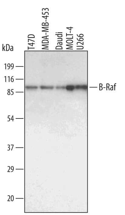
Detection of Human B-Raf by Western Blot.
Western blot shows lysates of T47D human breast cancer cell line, MDA-MB-453 human breast cancer cell line, Daudi human Burkitt's lymphoma cell line, MOLT-4 human acute lymphoblastic leukemia cell line and U266 human myeloma cell line. PVDF membrane was probed with 1 µg/mL of Goat Anti-Human B-Raf Antigen Affinity-purified Polyclonal Antibody (Catalog # AF3424) followed by HRP-conjugated Anti-Goat IgG Secondary Antibody (Catalog # HAF109). A specific band was detected for B-Raf at approximately 95 kDa (as indicated). This experiment was conducted under reducing conditions and using Immunoblot Buffer Group 1.Detection of Human B-Raf by Simple WesternTM.
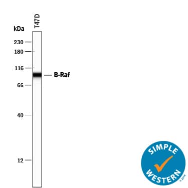
Simple Western lane view shows lysates of T47D human breast cancer cell line, loaded at 0.2 mg/mL. A specific band was detected for B-Raf at approximately 95 kDa (as indicated) using 10 µg/mL of Goat Anti-Human B-Raf Antigen Affinity-purified Polyclonal Antibody (Catalog # AF3424) followed by 1:50 dilution of HRP-conjugated Anti-Goat IgG Secondary Antibody (Catalog # HAF109). This experiment was conducted under reducing conditions and using the 12-230 kDa separation system.Detection of Human B-Raf by Simple WesternTM.
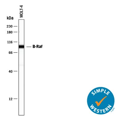
Simple Western lane view shows lysates of MOLT-4 human acute lymphoblastic leukemia cell line, loaded at 0.2 mg/mL. A specific band was detected for B-Raf at approximately 95 kDa (as indicated) using 10 µg/mL of Goat Anti-Human B-Raf Antigen Affinity-purified Polyclonal Antibody (Catalog # AF3424) followed by 1:50 dilution of HRP-conjugated Anti-Goat IgG Secondary Antibody (Catalog # HAF109). This experiment was conducted under reducing conditions and using the 12-230 kDa separation system.Applications for Human B-Raf Antibody
Simple Western
Sample: T47D human breast cancer cell line and MOLT‑4 human acute lymphoblastic leukemia cell line
Western Blot
Sample: T47D human breast cancer cell line, MDA-MB-453 human breast cancer cell line, Daudi human Burkitt's lymphoma cell line, MOLT-4 human acute lymphoblastic leukemia cell line and U266 human myeloma cell line
Reviewed Applications
Read 1 review rated 4 using AF3424 in the following applications:
Formulation, Preparation, and Storage
Purification
Reconstitution
Formulation
Shipping
Stability & Storage
- 12 months from date of receipt, -20 to -70 °C as supplied.
- 1 month, 2 to 8 °C under sterile conditions after reconstitution.
- 6 months, -20 to -70 °C under sterile conditions after reconstitution.
Background: B-Raf
The Raf serine/threonine kinases are effectors of Ras that function as MAP3Ks in the ERK phosphorylation cascade. Mammals express three Raf proteins: Raf-1 (C‑Raf); A-Raf; and B-Raf, found at high levels in cerebrum and testes. Mice with a targeted disruption of the B-Raf gene die of vascular defects during mid-gestation. B-Raf mutations have been found in two-thirds of malignant melanomas, with the single substitution V599E in the kinase domain the most frequent occurrence. B-Raf activation in melanomas results in BIM phosphorylation and inhibition of apoptosis.
Long Name
Alternate Names
Gene Symbol
UniProt
Additional B-Raf Products
Product Documents for Human B-Raf Antibody
Product Specific Notices for Human B-Raf Antibody
For research use only


