Human B7-2/CD86 APC-conjugated Antibody
R&D Systems, part of Bio-Techne | Catalog # FAB141A


Conjugate
Catalog #
Key Product Details
Validated by
Biological Validation
Species Reactivity
Validated:
Human
Cited:
Porcine
Applications
Validated:
Flow Cytometry
Cited:
Flow Cytometry
Label
Allophycocyanin (Excitation = 620-650 nm, Emission = 660-670 nm)
Antibody Source
Monoclonal Mouse IgG1 Clone # 37301
Product Specifications
Immunogen
S. frugiperda insect ovarian cell line Sf 21-derived recombinant human B7‑2/CD86
Specificity
Detects human B7‑2/CD86 in direct ELISAs and Western blots. In direct ELISAs, no cross-reactivity with recombinant human (rh) B7-1, recombinant mouse B7-2, recombinant rat B7-2, rhB7-H1, rhB7-H2, rhB7-H3, rhB7-H3b, rhB7-H4, or rhB7-L2 is observed.
Clonality
Monoclonal
Host
Mouse
Isotype
IgG1
Scientific Data Images for Human B7-2/CD86 APC-conjugated Antibody
Detection of B7‑2/CD86 in Human Blood Monocytes by Flow Cytometry.
Human peripheral blood monocytes were stained with Mouse Anti-Human CD14 PE-conjugated Monoclonal Antibody (Catalog # FAB3832P) and either (A) Mouse Anti-Human B7-2/CD86 APC-conjugated Monoclonal Antibody (Catalog # FAB141A) or (B) Mouse IgG1Allophycocyanin Isotype Control (Catalog # IC002A). View our protocol for Staining Membrane-associated Proteins.Detection of Porcine B7-2/CD86 by Flow Cytometry
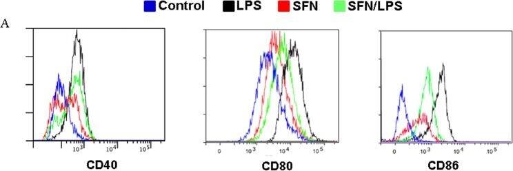
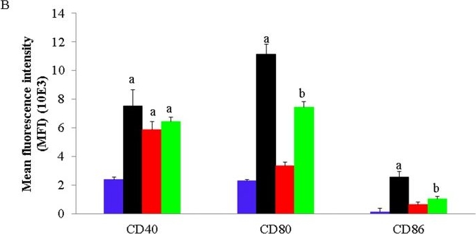
SFN inhibits LPS induced moDC maturation and enhances the phagocytic activity.moDCs at day 7 in culture were used for cell phagocytosis and cell differentiation status analysis. moDCs were pre-incubated for 1 h with or without SFN (10 μM) before stimulation for 24 h LPS (1.0 μg/ml) or to the indicated concentrations. CD40, CD80, and CD86 cellular surface markers expression were analyzed by flow cytometry (A). The flow cytometry results shown were from one experiment of two independent experiments. CD40, CD80 and CD86 mean fluorescence intensity (MFI) determined by flow cytometry (B). The flow cytometry results were combined from two independent experiments and each experiment was performed from triplications. Data are mean ± standard deviations (SD) (the letters a and b P<0.01). The phagocytic activity of moDCs was examined after stimulating with different concentration of LPS (0,5 μg/ml, 1,0 μg/ml, and 2,0 μg/ml) with or without 24 h pre-treatment with SFN (C). The mRNA expression of DCs surface markers CD40, CD80 and CD86 were quantified using qRT-PCR (D). The mRNA expression and phagocytosis results were combined from three independent experiments and each experiment was performed in four replications. The data represented as the mean ± standard deviations (SD) (* P < 0.05; ** P < 0.01; *** P < 0.001). Image collected and cropped by CiteAb from the following publication (https://pubmed.ncbi.nlm.nih.gov/25793534), licensed under a CC-BY license. Not internally tested by R&D Systems.Detection of Porcine B7-2/CD86 by Flow Cytometry
SFN inhibits LPS induced moDC maturation and enhances the phagocytic activity.moDCs at day 7 in culture were used for cell phagocytosis and cell differentiation status analysis. moDCs were pre-incubated for 1 h with or without SFN (10 μM) before stimulation for 24 h LPS (1.0 μg/ml) or to the indicated concentrations. CD40, CD80, and CD86 cellular surface markers expression were analyzed by flow cytometry (A). The flow cytometry results shown were from one experiment of two independent experiments. CD40, CD80 and CD86 mean fluorescence intensity (MFI) determined by flow cytometry (B). The flow cytometry results were combined from two independent experiments and each experiment was performed from triplications. Data are mean ± standard deviations (SD) (the letters a and b P<0.01). The phagocytic activity of moDCs was examined after stimulating with different concentration of LPS (0,5 μg/ml, 1,0 μg/ml, and 2,0 μg/ml) with or without 24 h pre-treatment with SFN (C). The mRNA expression of DCs surface markers CD40, CD80 and CD86 were quantified using qRT-PCR (D). The mRNA expression and phagocytosis results were combined from three independent experiments and each experiment was performed in four replications. The data represented as the mean ± standard deviations (SD) (* P < 0.05; ** P < 0.01; *** P < 0.001). Image collected and cropped by CiteAb from the following publication (https://pubmed.ncbi.nlm.nih.gov/25793534), licensed under a CC-BY license. Not internally tested by R&D Systems.Applications for Human B7-2/CD86 APC-conjugated Antibody
Application
Recommended Usage
Flow Cytometry
10 µL/106 cells
Sample: Human peripheral blood monocytes
Sample: Human peripheral blood monocytes
Reviewed Applications
Read 1 review rated 5 using FAB141A in the following applications:
Formulation, Preparation, and Storage
Purification
Protein A or G purified from ascites
Formulation
Supplied in a saline solution containing BSA and Sodium Azide.
Shipping
The product is shipped with polar packs. Upon receipt, store it immediately at the temperature recommended below.
Stability & Storage
Protect from light. Do not freeze.
- 12 months from date of receipt, 2 to 8 °C as supplied.
Background: B7-2/CD86
References
- Azuma, M. et al. (1993) Nature 366:76.
- Freeman, G.J. et al. (1993) Science 262:909.
- Freeman, G. et al. (1991) J. Exp. Med. 174:625.
- Selvakumar, A. et al. (1993) Immunogenetics 38:292.
- Chen, C. et al. (1994) J. Immunol. 152:4929.
- Freeman, G.J. et al. (1993) J. Exp. Med. 178:2185.
Alternate Names
B72, CD86
Gene Symbol
CD86
Additional B7-2/CD86 Products
Product Documents for Human B7-2/CD86 APC-conjugated Antibody
Product Specific Notices for Human B7-2/CD86 APC-conjugated Antibody
For research use only
Loading...
Loading...
Loading...
Loading...
Loading...

