Human CaM Kinase I Antibody
R&D Systems, part of Bio-Techne | Catalog # AF7899

Key Product Details
Species Reactivity
Applications
Label
Antibody Source
Product Specifications
Immunogen
Ser176-Arg254 (Ala224Ser)
Accession # Q14012
Specificity
Clonality
Host
Isotype
Scientific Data Images for Human CaM Kinase I Antibody
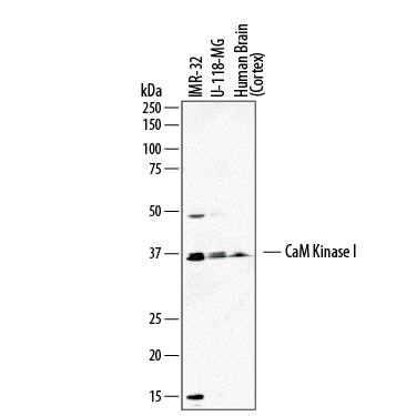
Detection of Human CaM Kinase I by Western Blot.
Western blot shows lysates of IMR-32 human neuroblastoma cell line, U-118-MG human glioblastoma/astrocytoma cell line, and human brain (cortex) tissue. PVDF membrane was probed with 2 µg/mL of Sheep Anti-Human CaM Kinase I Antigen Affinity-purified Polyclonal Antibody (Catalog # AF7899) followed by HRP-conjugated Anti-Sheep IgG Secondary Antibody (Catalog # HAF016). A specific band was detected for CaM Kinase I at approximately 37 kDa (as indicated). This experiment was conducted under reducing conditions and using Immunoblot Buffer Group 1.CaM Kinase I alpha in Human Hypothalamus.
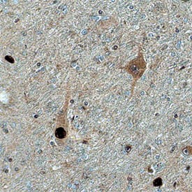
CaM Kinase I was detected in immersion fixed paraffin-embedded sections of human hypothalamus using Sheep Anti-Human CaM Kinase I Antigen Affinity-purified Polyclonal Antibody (Catalog # AF7899) at 1.7 µg/mL overnight at 4 °C. Tissue was stained using the Anti-Sheep HRP-DAB Cell & Tissue Staining Kit (brown; Catalog # CTS019) and counterstained with hematoxylin (blue). Specific staining was localized to cytoplasm and nuclei. View our protocol for Chromogenic IHC Staining of Paraffin-embedded Tissue Sections.Applications for Human CaM Kinase I Antibody
Immunohistochemistry
Sample: Immersion fixed paraffin-embedded sections of human hypothalamus
Western Blot
Sample: IMR‑32 human neuroblastoma cell line, U‑118‑MG human glioblastoma/astrocytoma cell line, and human brain (cortex) tissue
Formulation, Preparation, and Storage
Purification
Reconstitution
Formulation
Shipping
Stability & Storage
- 12 months from date of receipt, -20 to -70 °C as supplied.
- 1 month, 2 to 8 °C under sterile conditions after reconstitution.
- 6 months, -20 to -70 °C under sterile conditions after reconstitution.
Background: CaM Kinase I
CaMKI (Calcium/Calmodulin-dependent kinase I) is a 37-43 kDa monomeric and cytosolic member of the CaMK subfamily, Ser/Thr protein kinase family, protein kinase superfamily of molecules. It is found in multiple cell types, including adrenal cortical cells, neurons and macrophages/osteoclast precursors. CaMKI is associated with the cellular Ca++ signaling pathway. Upon entry into the cell, calcium levels are sensed by calmodulin. Upon Ca++ binding, calmodulin (and CaMK kinase) interacts with a Ser/Thr protein kinase termed CaMKIa. CaMKIa is of particular interest in neurons and cells of the zona glomerulosa. In neurons, CaMKIa activation results in the recruitment of AMPA-Rs and the promotion of axon growth (vs.dendrite outgrowth which is promoted by CaMKIg). In the adrenal gland, CaMKIa regulates CYP11B2 transcription, and thus the ability to synthesize aldosterone. Human CaMKI, the isoform used for immunization, is 370 amino acids (aa) in length (SwissProt #:Q14012). It contains one protein kinase domain (aa 20-276) followed by a calmodulin-binding region (aa 296-317) and an NES (aa 315-321). Over aa 178-257, human and mouse CaMKI are identical in amino acid sequence. There are three additional CaMKI isoforms (b, g, d), all the product of distinct genes. Nevertheless, over the same aa sequence, these three isoforms (b, g, d) share 80%, 81% and 97% aa sequence identity with CaMKI(a), respectively.
Long Name
Alternate Names
UniProt
Additional CaM Kinase I Products
Product Documents for Human CaM Kinase I Antibody
Product Specific Notices for Human CaM Kinase I Antibody
For research use only

